Copyright
©The Author(s) 2019.
World J Clin Cases. Apr 26, 2019; 7(8): 992-1000
Published online Apr 26, 2019. doi: 10.12998/wjcc.v7.i8.992
Published online Apr 26, 2019. doi: 10.12998/wjcc.v7.i8.992
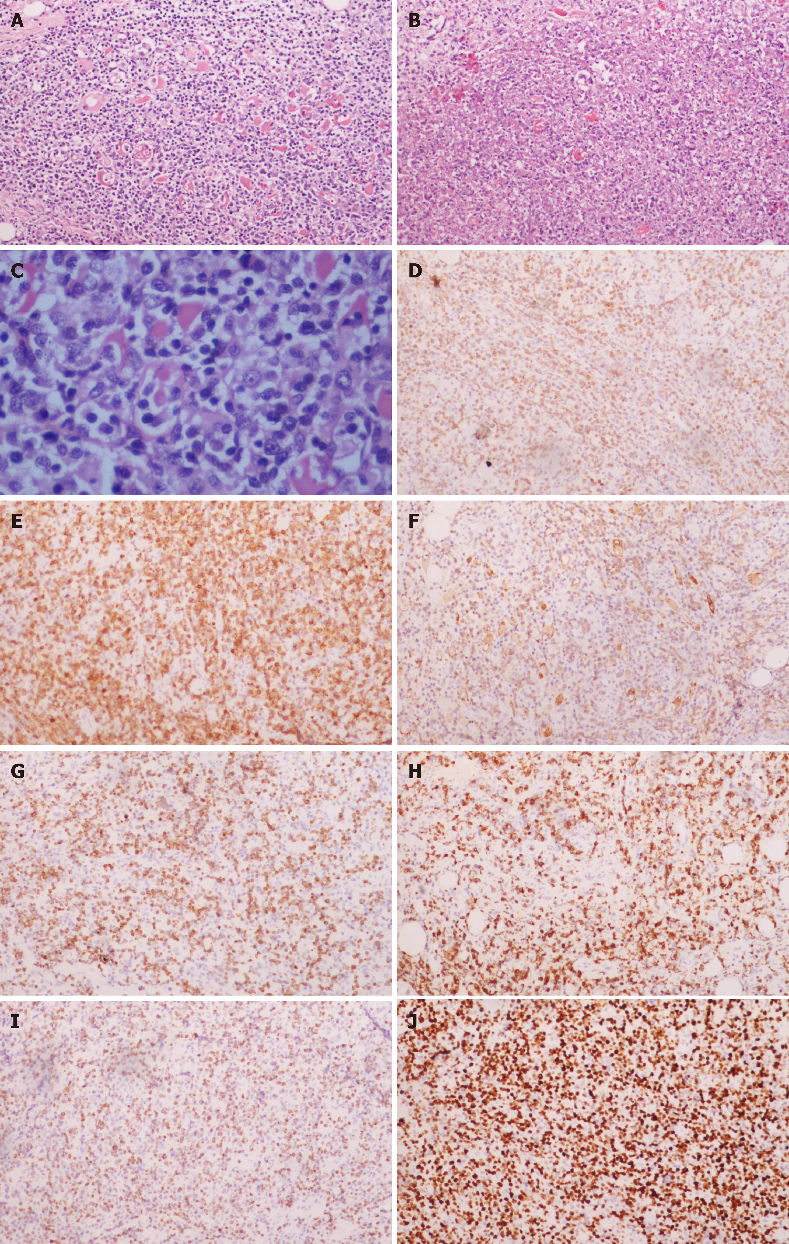
Figure 4 Histopathological and immunohistochemical examinations.
A-C: Hematoxylin-eosin staining of tumor cells (A, 100× magnification, C, 400× magnification) and tumor-associated necrotic tissue (B, 100× magnification); D-J: immunohistochemical staining for CD2 (D), CD3 (E), CD56 (F), perforin (G), granzyme B (H), TIA-1 (I), and Ki-67 (J).
- Citation: Liu YN, Zhu Y, Tan JJ, Shen GS, Huang SL, Zhou CG, Huangfu SH, Zhang R, Huang XB, Wang L, Zhang Q, Jiang B. Extranodal natural killer/T-cell lymphoma (nasal type) presenting as a perianal abscess: A case report. World J Clin Cases 2019; 7(8): 992-1000
- URL: https://www.wjgnet.com/2307-8960/full/v7/i8/992.htm
- DOI: https://dx.doi.org/10.12998/wjcc.v7.i8.992


