Copyright
©The Author(s) 2019.
World J Clin Cases. Apr 26, 2019; 7(8): 984-991
Published online Apr 26, 2019. doi: 10.12998/wjcc.v7.i8.984
Published online Apr 26, 2019. doi: 10.12998/wjcc.v7.i8.984
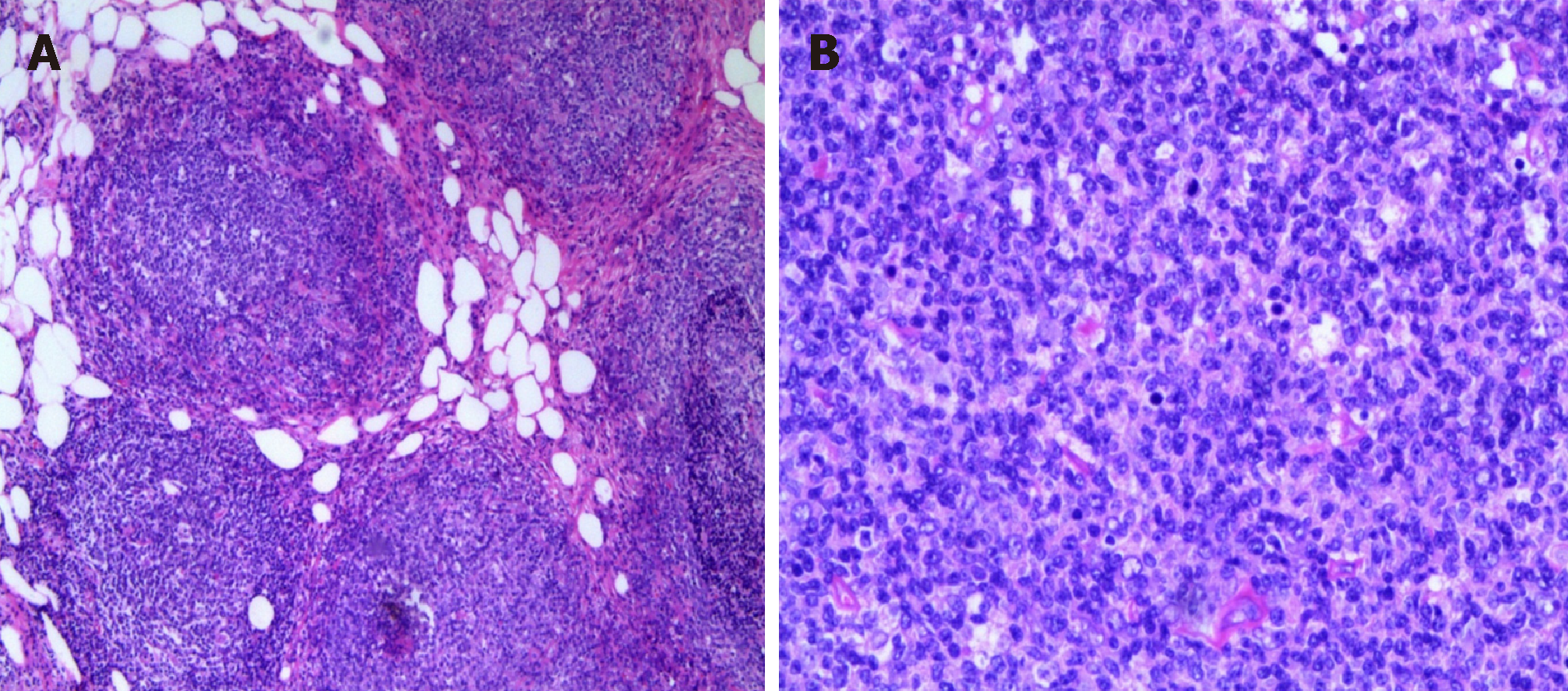
Figure 4 Omental histopathological examination.
A: Lymphoid hyperplasia in omental adipose tissue; B: Some follicles were fused, and no typical cuff was observed. The follicular node was seen under a light microscope (magnification, 200×). The section consists of central cells and centroblasts, and the number of centroblasts is > 15/HPF.
- Citation: Wei C, Xiong F, Yu ZC, Li DF, Luo MH, Liu TT, Li YX, Zhang DG, Xu ZL, Jin HT, Tang Q, Wang LS, Wang JY, Yao J. Diagnosis of follicular lymphoma by laparoscopy: A case report. World J Clin Cases 2019; 7(8): 984-991
- URL: https://www.wjgnet.com/2307-8960/full/v7/i8/984.htm
- DOI: https://dx.doi.org/10.12998/wjcc.v7.i8.984


