Copyright
©The Author(s) 2019.
World J Clin Cases. Apr 26, 2019; 7(8): 984-991
Published online Apr 26, 2019. doi: 10.12998/wjcc.v7.i8.984
Published online Apr 26, 2019. doi: 10.12998/wjcc.v7.i8.984
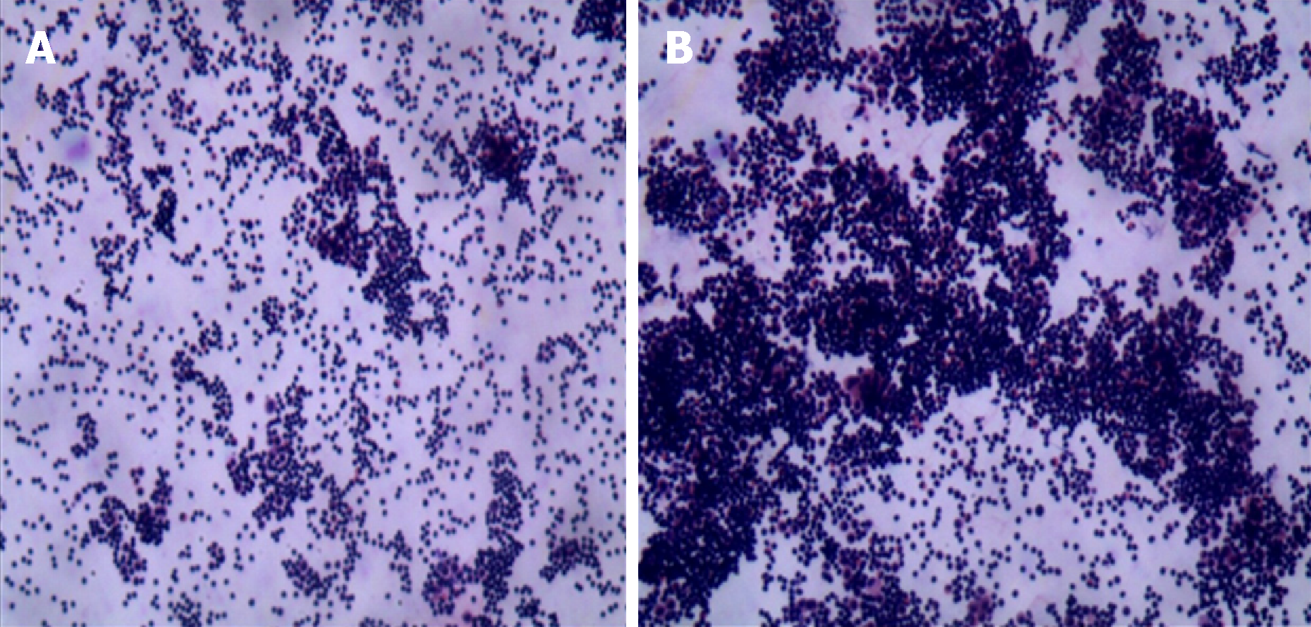
Figure 3 Cytology results of ascites.
A: A large number of lymphocytes can be seen under a light microscope (magnification, 100×); B: A large number of lymphocytes are visible under a light microscope (magnification, 200×). The mesothelial cells, phagocytes, and a few eosinophils can be seen.
- Citation: Wei C, Xiong F, Yu ZC, Li DF, Luo MH, Liu TT, Li YX, Zhang DG, Xu ZL, Jin HT, Tang Q, Wang LS, Wang JY, Yao J. Diagnosis of follicular lymphoma by laparoscopy: A case report. World J Clin Cases 2019; 7(8): 984-991
- URL: https://www.wjgnet.com/2307-8960/full/v7/i8/984.htm
- DOI: https://dx.doi.org/10.12998/wjcc.v7.i8.984


