Copyright
©The Author(s) 2019.
World J Clin Cases. Apr 26, 2019; 7(8): 972-983
Published online Apr 26, 2019. doi: 10.12998/wjcc.v7.i8.972
Published online Apr 26, 2019. doi: 10.12998/wjcc.v7.i8.972
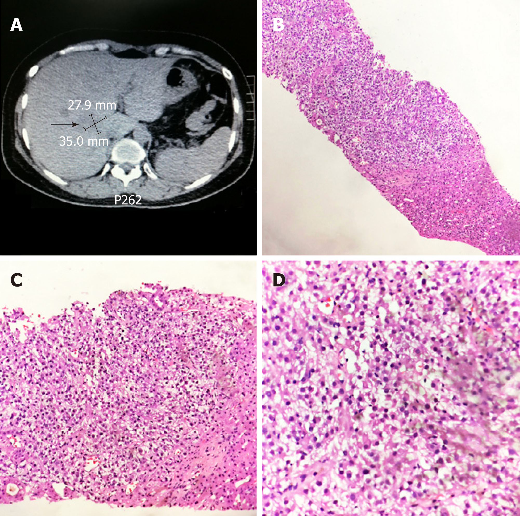
Figure 2 Computed tomography guided percutaneous liver tumor puncture and pathological findings of case 2.
A: Computed tomography scan revealed a mass of 3.5 cm × 3.0 cm in liver; B-D: The pathological diagnosis suggested metastatic hepatic epithelioid angiomyolipoma (HE staining, B: × 100, C: × 200, D: × 400).
- Citation: Mao JX, Teng F, Liu C, Yuan H, Sun KY, Zou Y, Dong JY, Ji JS, Dong JF, Fu H, Ding GS, Guo WY. Two case reports and literature review for hepatic epithelioid angiomyolipoma: Pitfall of misdiagnosis. World J Clin Cases 2019; 7(8): 972-983
- URL: https://www.wjgnet.com/2307-8960/full/v7/i8/972.htm
- DOI: https://dx.doi.org/10.12998/wjcc.v7.i8.972


