Copyright
©The Author(s) 2019.
World J Clin Cases. Apr 26, 2019; 7(8): 972-983
Published online Apr 26, 2019. doi: 10.12998/wjcc.v7.i8.972
Published online Apr 26, 2019. doi: 10.12998/wjcc.v7.i8.972
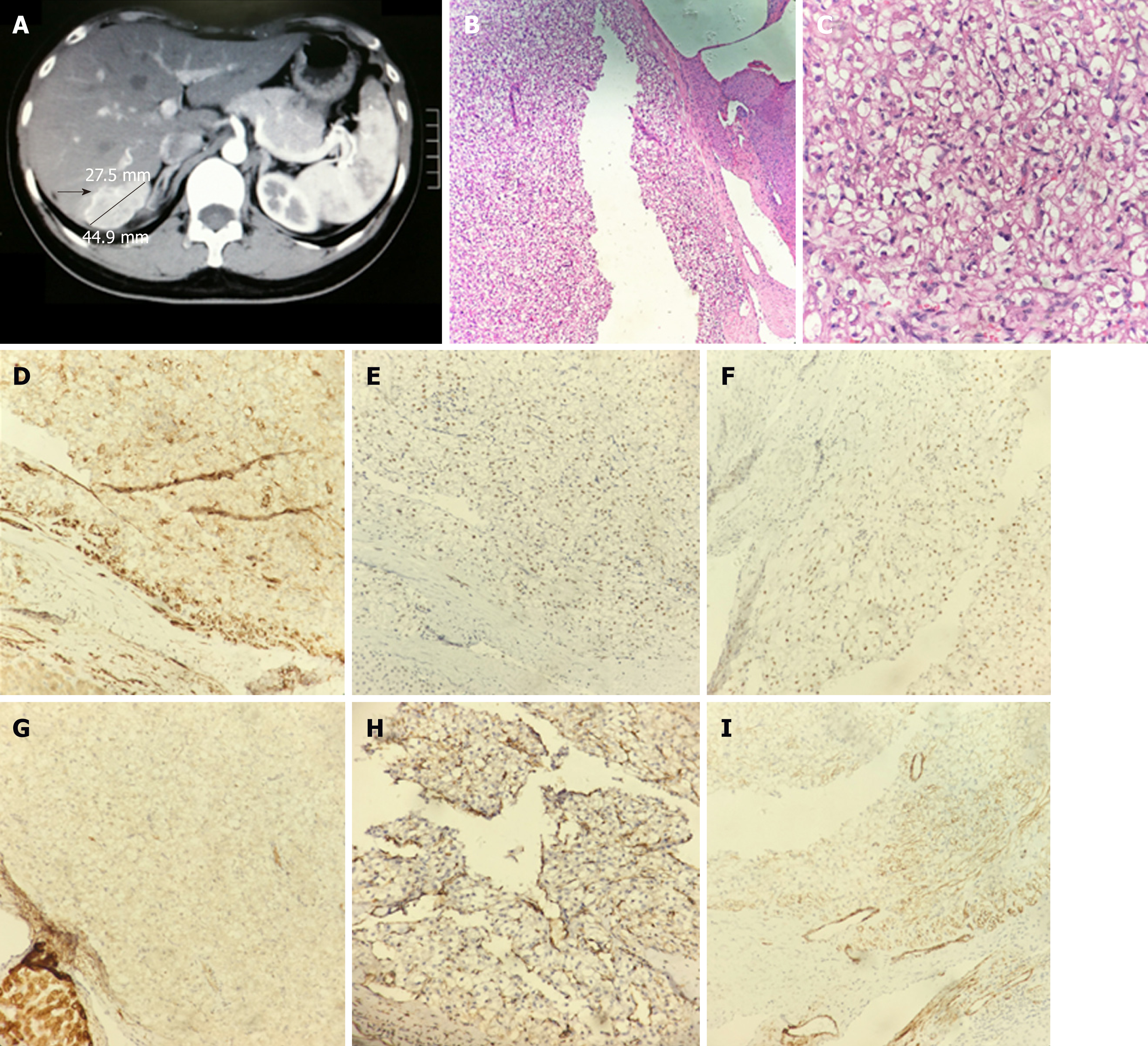
Figure 1 Imaging and pathological immunohistochemical findings of case 1.
A: Computed tomography scan suggested a mass of 5 cm × 3 cm in the right lobe of liver; B, C: The postoperative pathological diagnosis was hepatic epithelioid angiomyolipoma (HE staining, B: × 100, C: × 400); D-I (× 200): Immunohistochemistry results were as follows: Calponin (++) (D); estrogen receptor (positive in some cells) (E); progesterone receptor (+) (F); K-pan (weak positive) (G); vimentin (partially positive) (H); and actin (partially positive) (I). HE: Hematoxylin and eosin.
- Citation: Mao JX, Teng F, Liu C, Yuan H, Sun KY, Zou Y, Dong JY, Ji JS, Dong JF, Fu H, Ding GS, Guo WY. Two case reports and literature review for hepatic epithelioid angiomyolipoma: Pitfall of misdiagnosis. World J Clin Cases 2019; 7(8): 972-983
- URL: https://www.wjgnet.com/2307-8960/full/v7/i8/972.htm
- DOI: https://dx.doi.org/10.12998/wjcc.v7.i8.972


