Copyright
©The Author(s) 2019.
World J Clin Cases. Apr 6, 2019; 7(7): 872-880
Published online Apr 6, 2019. doi: 10.12998/wjcc.v7.i7.872
Published online Apr 6, 2019. doi: 10.12998/wjcc.v7.i7.872
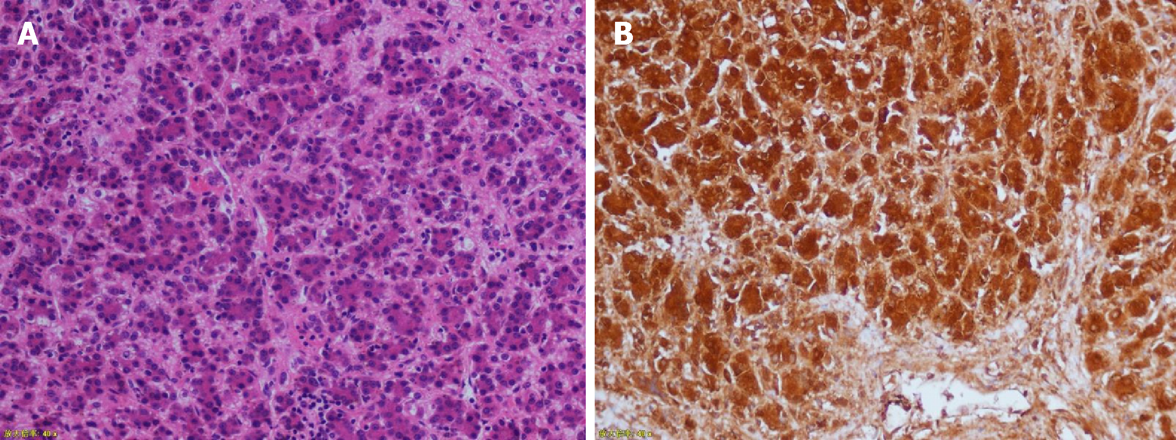
Figure 6 Pathological images (magnification 200×).
The immunohistochemical results were hepatocytes (+), GPC-3 (+), alpha fetoprotein (focal +), ki-67 (+20%), Arg-1 (+), and CD99 (local).
- Citation: Chen DX, Wang SJ, Jiang YN, Yu MC, Fan JZ, Wang XQ. Robot-assisted gallbladder-preserving hepatectomy for treating S5 hepatoblastoma in a child: A case report and review of the literature. World J Clin Cases 2019; 7(7): 872-880
- URL: https://www.wjgnet.com/2307-8960/full/v7/i7/872.htm
- DOI: https://dx.doi.org/10.12998/wjcc.v7.i7.872


