Copyright
©The Author(s) 2019.
World J Clin Cases. Apr 6, 2019; 7(7): 863-871
Published online Apr 6, 2019. doi: 10.12998/wjcc.v7.i7.863
Published online Apr 6, 2019. doi: 10.12998/wjcc.v7.i7.863
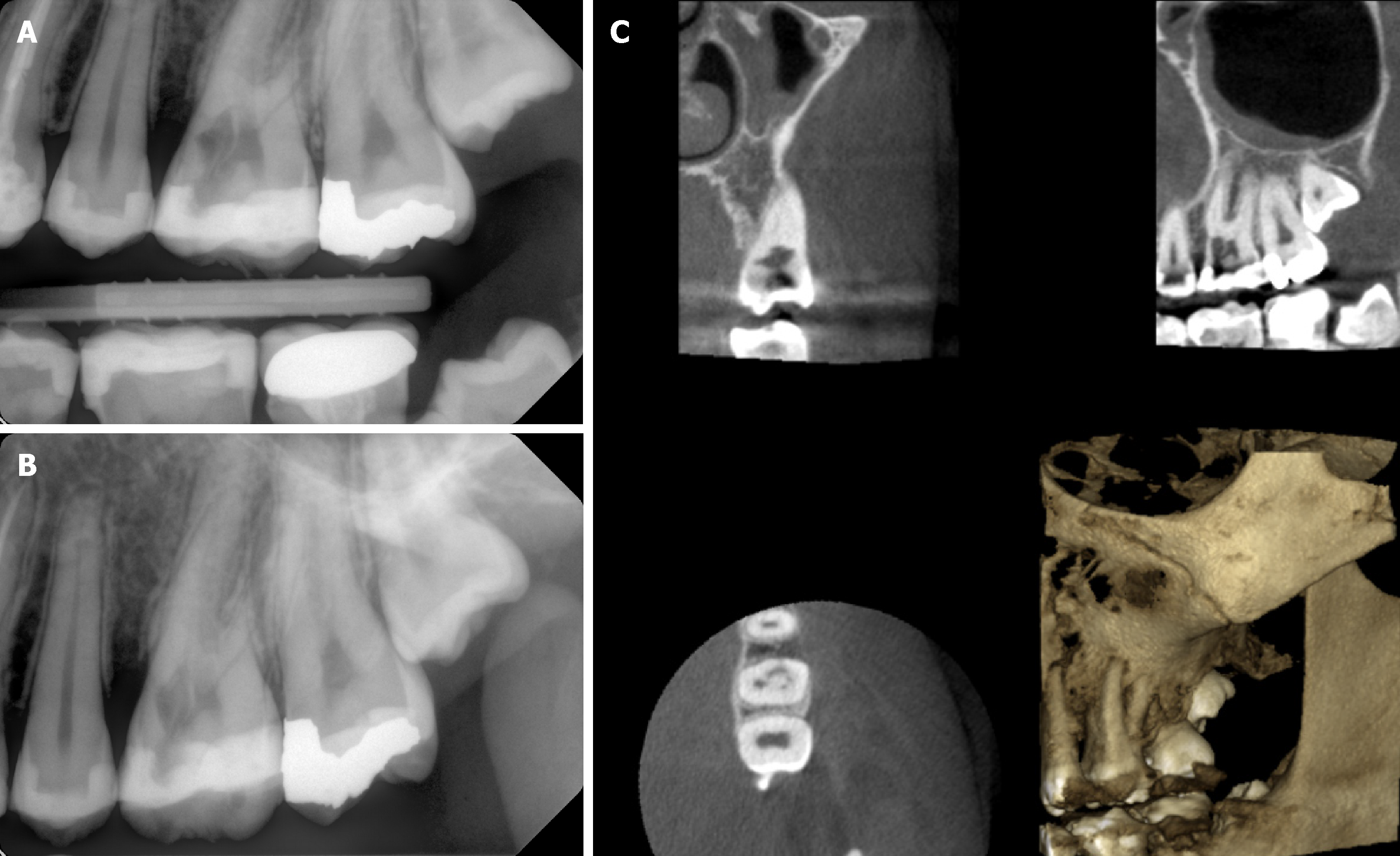
Figure 2 Radiographic findings.
A, B: Two dimensional radiographs revealed breakdown of alveolar crest mesial to the left maxillary first molar and irregular molted radiolucency mesial to the pulp space of the left maxillary first molar separated by radiopaque line and extending into the radicular dentin; C: Cone-beam computed tomographic images from different planes demonstrated the size and external perforation of the resorption defect.
- Citation: Alqedairi A. Non-Invasive management of invasive cervical resorption associated with periodontal pocket: A case report. World J Clin Cases 2019; 7(7): 863-871
- URL: https://www.wjgnet.com/2307-8960/full/v7/i7/863.htm
- DOI: https://dx.doi.org/10.12998/wjcc.v7.i7.863


