Copyright
©The Author(s) 2019.
World J Clin Cases. Apr 6, 2019; 7(7): 855-862
Published online Apr 6, 2019. doi: 10.12998/wjcc.v7.i7.855
Published online Apr 6, 2019. doi: 10.12998/wjcc.v7.i7.855
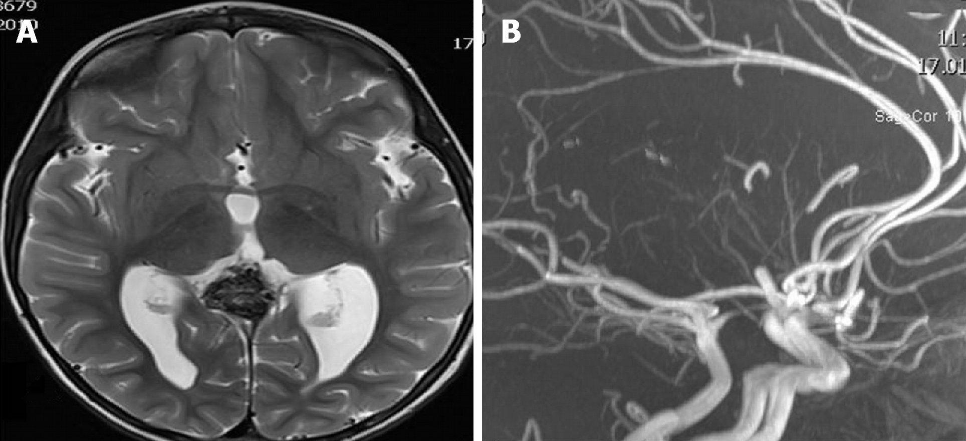
Figure 3 Imaging examinations of patient 3.
A: The MR angiography one year after treatment showed mild ventricular dilatation, especially of the left occipital horn; B: No residual flow in the VGAM is seen. The VGAM was occluded completely.
- Citation: Spazzapan P, Milosevic Z, Velnar T. Vein of Galen aneurismal malformations - clinical characteristics, treatment and presentation: Three cases report. World J Clin Cases 2019; 7(7): 855-862
- URL: https://www.wjgnet.com/2307-8960/full/v7/i7/855.htm
- DOI: https://dx.doi.org/10.12998/wjcc.v7.i7.855


