Copyright
©The Author(s) 2019.
World J Clin Cases. Mar 26, 2019; 7(6): 785-791
Published online Mar 26, 2019. doi: 10.12998/wjcc.v7.i6.785
Published online Mar 26, 2019. doi: 10.12998/wjcc.v7.i6.785
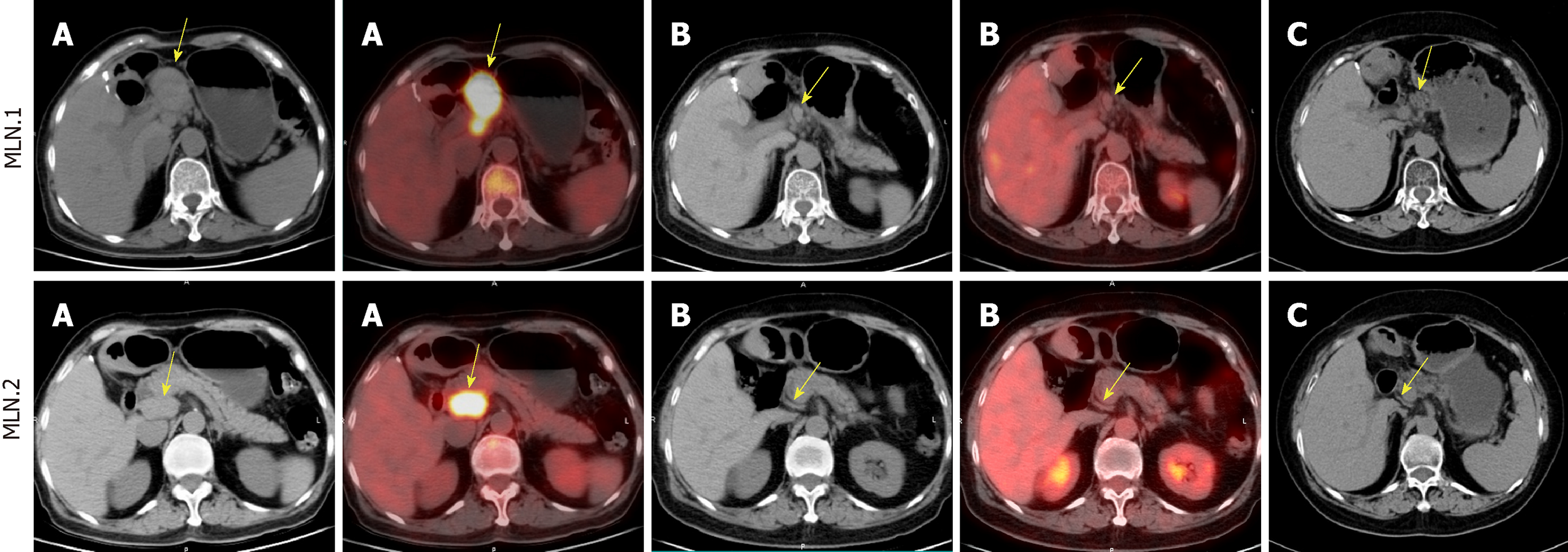
Figure 1 Computed tomography or fluorine-18-Fluorodeoxyglucose positron emission tomography/computed tomography images of metastatic lymph nodes.
A: Baseline image at progression after surgery; B: After eight cycles of chemotherapy with gemcitabine and docetaxel; C: Computed tomography image one month after radiotherapy. MLN.1: Metastatic lymph nodes in the hepatogastric ligament; MLN.2: Metastatic lymph nodes in the portacaval space in the abdomen are showed in chronological order (arrow).
- Citation: Chen HM, Shen YL, Liu M. Primary hepatic follicular dendritic cell sarcoma: A case report. World J Clin Cases 2019; 7(6): 785-791
- URL: https://www.wjgnet.com/2307-8960/full/v7/i6/785.htm
- DOI: https://dx.doi.org/10.12998/wjcc.v7.i6.785


