Copyright
©The Author(s) 2019.
World J Clin Cases. Mar 26, 2019; 7(6): 753-758
Published online Mar 26, 2019. doi: 10.12998/wjcc.v7.i6.753
Published online Mar 26, 2019. doi: 10.12998/wjcc.v7.i6.753
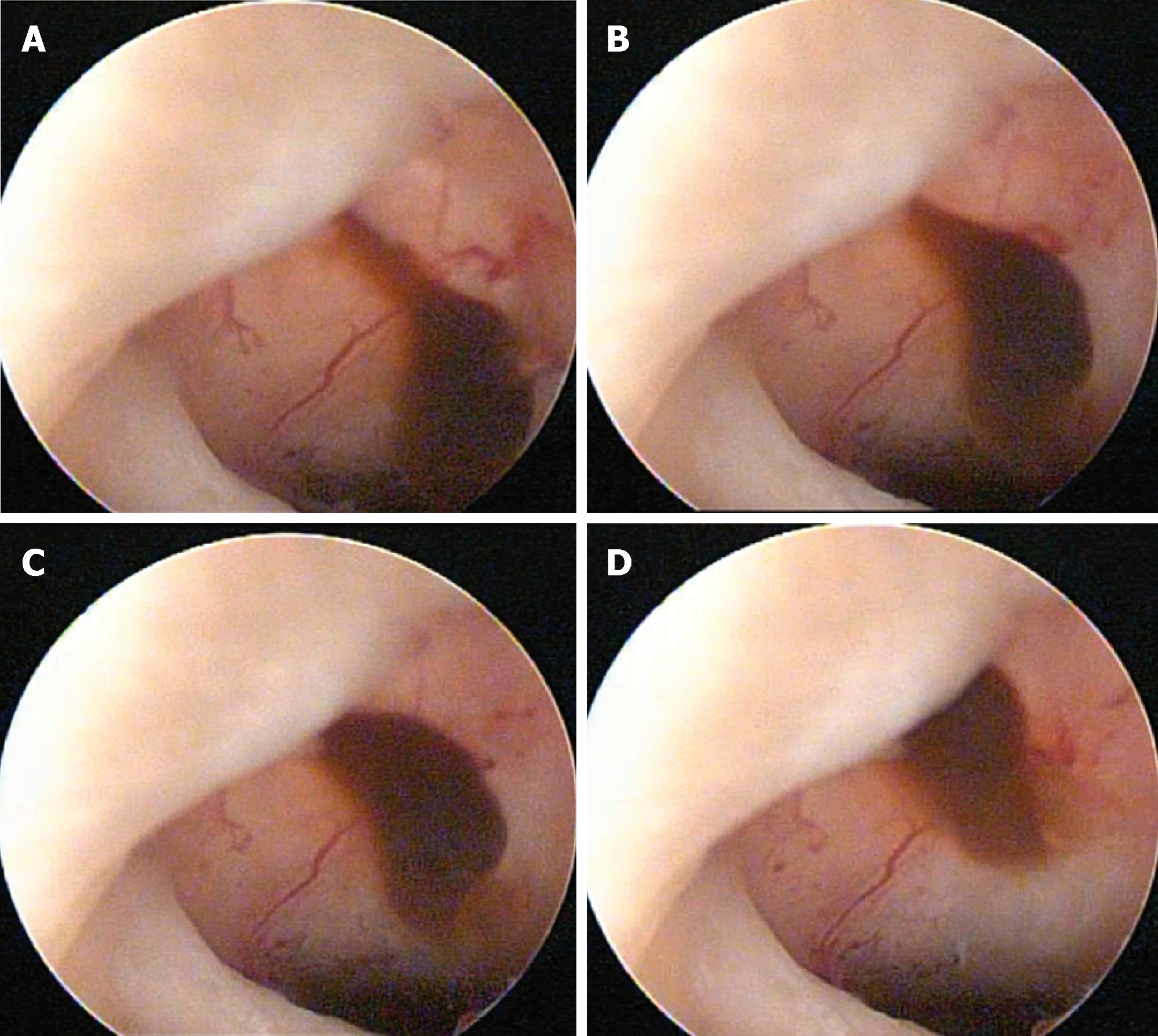
Figure 2 Isthmocele at the time of surgery.
Images were selected from the video recorded during surgery. The procedure was performed by finding the myocell sac containing neovascularization and mucosanguineous content, leveling the isthmocele area with a monopolar resectoscope loop, and subsequent application of monopolar ablation energy in the bed of the isthmocele using a roll ball electrode. The procedure was performed guided at times by transabdominal ultrasound (procedure time: 25 min; 2× magnification).
- Citation: López Rivero LP, Jaimes M, Camargo F, López-Bayghen E. Successful treatment with hysteroscopy for infertility due to isthmocele and hydrometra secondary to cesarean section: A case report. World J Clin Cases 2019; 7(6): 753-758
- URL: https://www.wjgnet.com/2307-8960/full/v7/i6/753.htm
- DOI: https://dx.doi.org/10.12998/wjcc.v7.i6.753


