Copyright
©The Author(s) 2019.
World J Clin Cases. Mar 26, 2019; 7(6): 753-758
Published online Mar 26, 2019. doi: 10.12998/wjcc.v7.i6.753
Published online Mar 26, 2019. doi: 10.12998/wjcc.v7.i6.753
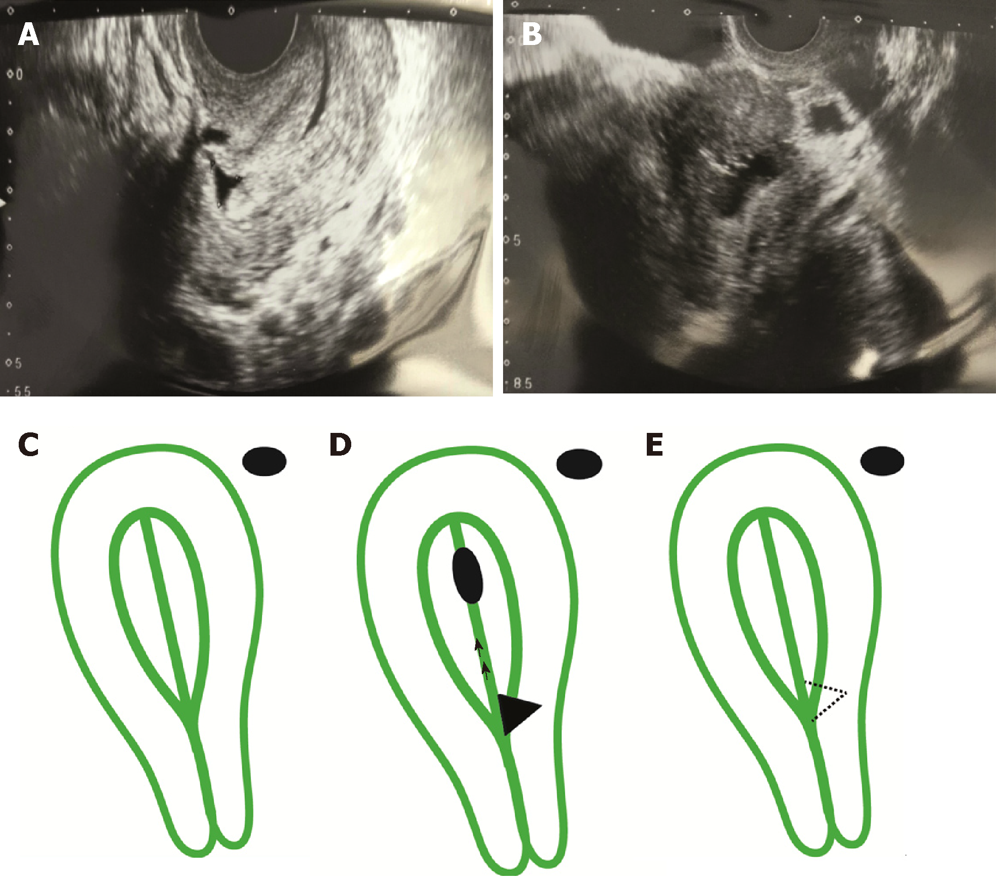
Figure 1 Transvaginal ultrasonographic images.
A 37-year-old female suffering from persistent hydrometra during in vitro fertilization underwent a transvaginal ultrasonographic examination. A: Panel A indicates the presence of a second-grade isthmocele; size 6.6 mm (base) × 6.1 mm (height); B: Panel B demonstrates the hydrometra and isthmocele. All images were taken with a Toshiba Xario 100 ultrasound with an endovaginal probe, frequency 7 megahertz (MHz); C: Diagrammatic representations of the uterus showing a normal uterus in Panel C; D: Hydrometra and isthmocele in Panel D; E: and result after resection in Panel E.
- Citation: López Rivero LP, Jaimes M, Camargo F, López-Bayghen E. Successful treatment with hysteroscopy for infertility due to isthmocele and hydrometra secondary to cesarean section: A case report. World J Clin Cases 2019; 7(6): 753-758
- URL: https://www.wjgnet.com/2307-8960/full/v7/i6/753.htm
- DOI: https://dx.doi.org/10.12998/wjcc.v7.i6.753


