Copyright
©The Author(s) 2019.
World J Clin Cases. Mar 26, 2019; 7(6): 717-726
Published online Mar 26, 2019. doi: 10.12998/wjcc.v7.i6.717
Published online Mar 26, 2019. doi: 10.12998/wjcc.v7.i6.717
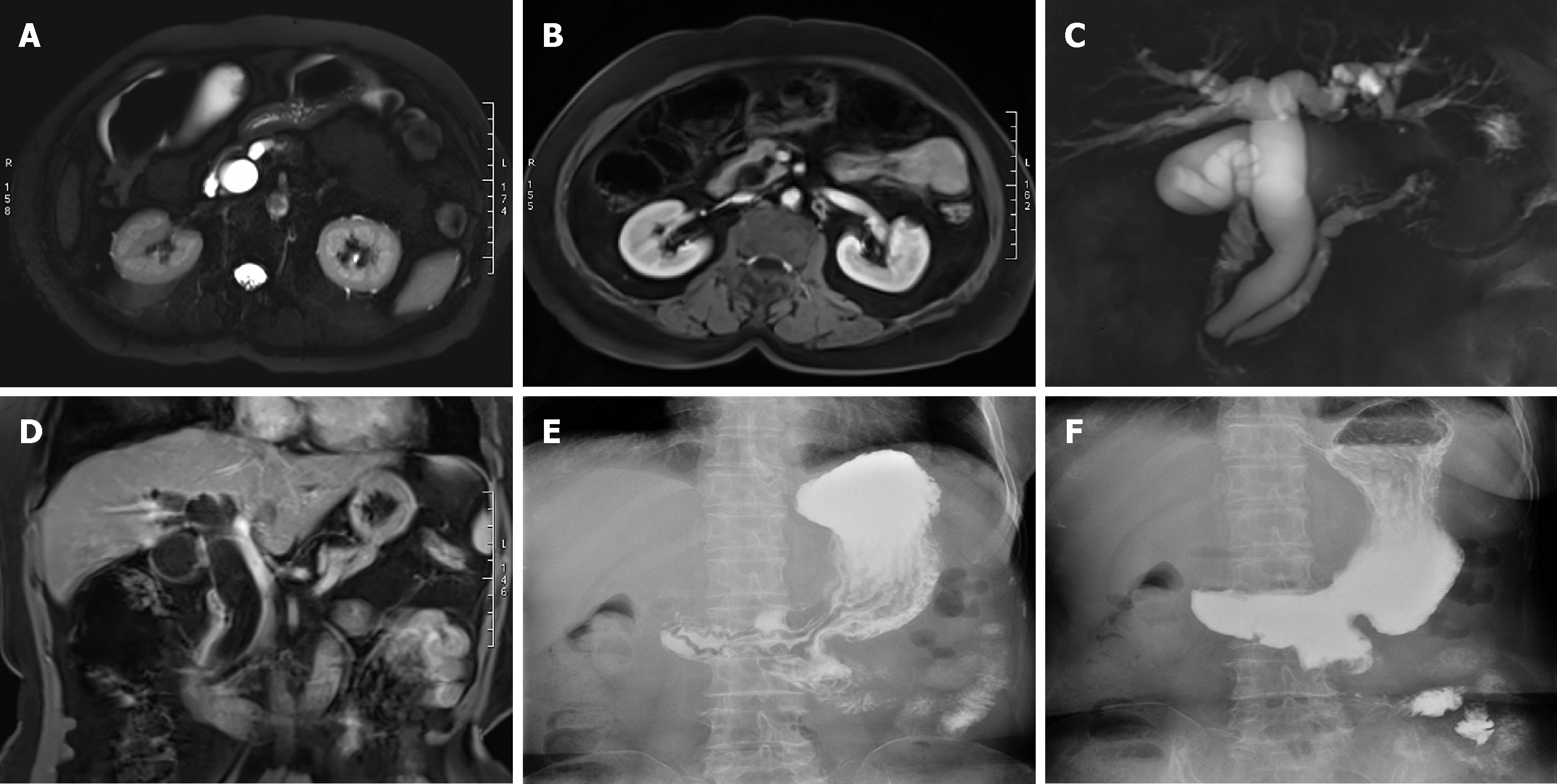
Figure 2 Magnetic resonance imaging and upper gastrointestinal iodine oil radiography.
A: Magnetic resonance imaging (MRI) T2-weighted imaging (T2WI) showing bile duct and pancreatic duct expansion; B: Enhanced MRI T1-weighted imaging (T1WI) showing bile duct and pancreatic duct expansion; C: Magnetic resonance cholangiopancreatography showing bile duct and pancreatic duct expansion and separation in the ampulla; D: Coronal MRI T1WI showing bile duct and pancreatic duct expansion; E: Upper gastrointestinal iodine oil radiography 6 mo after operation; gastric jejunum anastomosis showed stronger gastric emptying function; F: Common bile duct and pancreatic duct nonvisualization; no bile reflux in different phases.
- Citation: Liu F, Cheng JL, Cui J, Xu ZZ, Fu Z, Liu J, Tian H. Surgical method choice and coincidence rate of pathological diagnoses in transduodenal ampullectomy: A retrospective case series study and review of the literature. World J Clin Cases 2019; 7(6): 717-726
- URL: https://www.wjgnet.com/2307-8960/full/v7/i6/717.htm
- DOI: https://dx.doi.org/10.12998/wjcc.v7.i6.717


