Copyright
©The Author(s) 2019.
World J Clin Cases. Mar 6, 2019; 7(5): 676-683
Published online Mar 6, 2019. doi: 10.12998/wjcc.v7.i5.676
Published online Mar 6, 2019. doi: 10.12998/wjcc.v7.i5.676
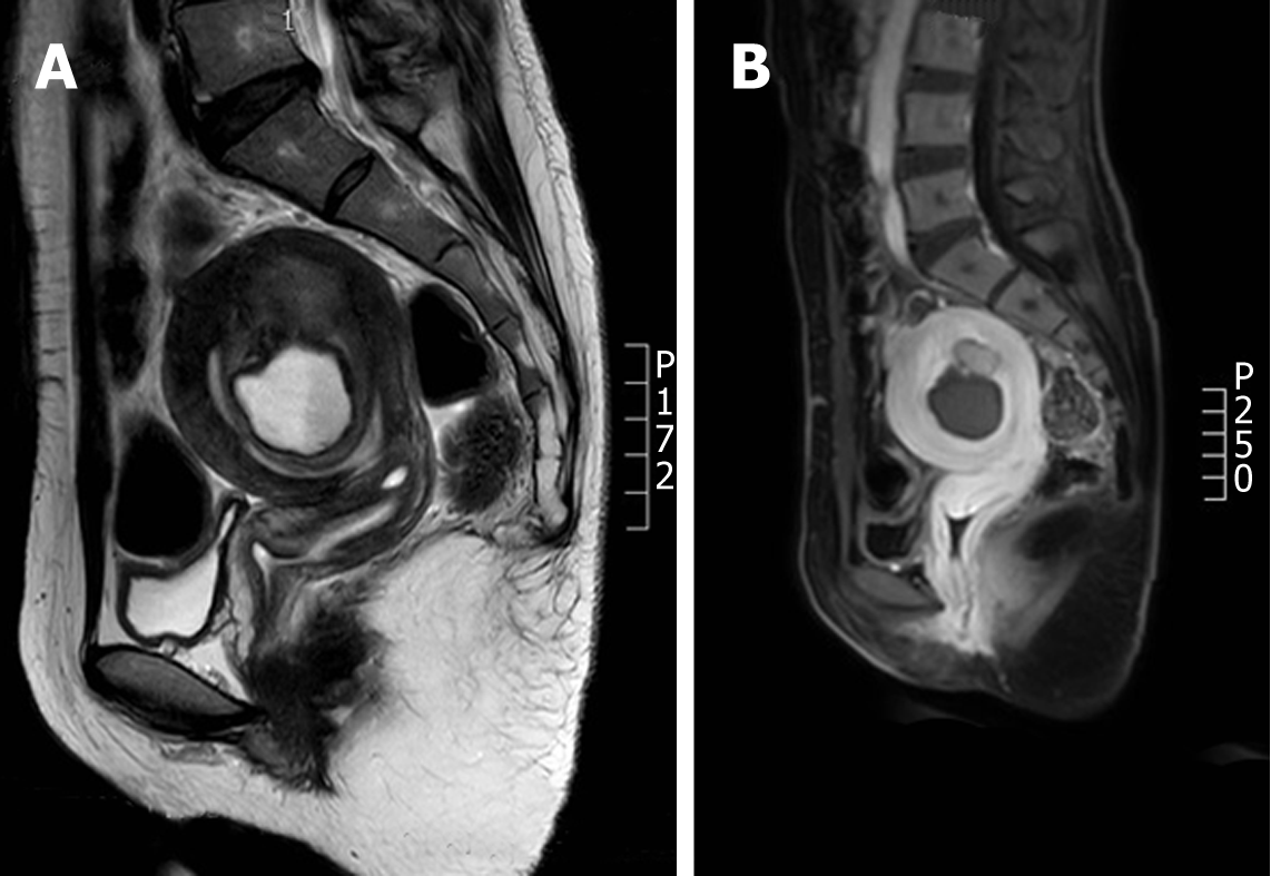
Figure 3 Magnetic resonance images of cystic adenomyosis (hyperintensity in T1 weighted image, moderate to high intensity in T2 weighted image, and low intensity in the edge).
A: Case 1; B: Case 2.
- Citation: Fan YY, Liu YN, Li J, Fu Y. Intrauterine cystic adenomyosis: Report of two cases. World J Clin Cases 2019; 7(5): 676-683
- URL: https://www.wjgnet.com/2307-8960/full/v7/i5/676.htm
- DOI: https://dx.doi.org/10.12998/wjcc.v7.i5.676


