Copyright
©The Author(s) 2019.
World J Clin Cases. Mar 6, 2019; 7(5): 628-635
Published online Mar 6, 2019. doi: 10.12998/wjcc.v7.i5.628
Published online Mar 6, 2019. doi: 10.12998/wjcc.v7.i5.628
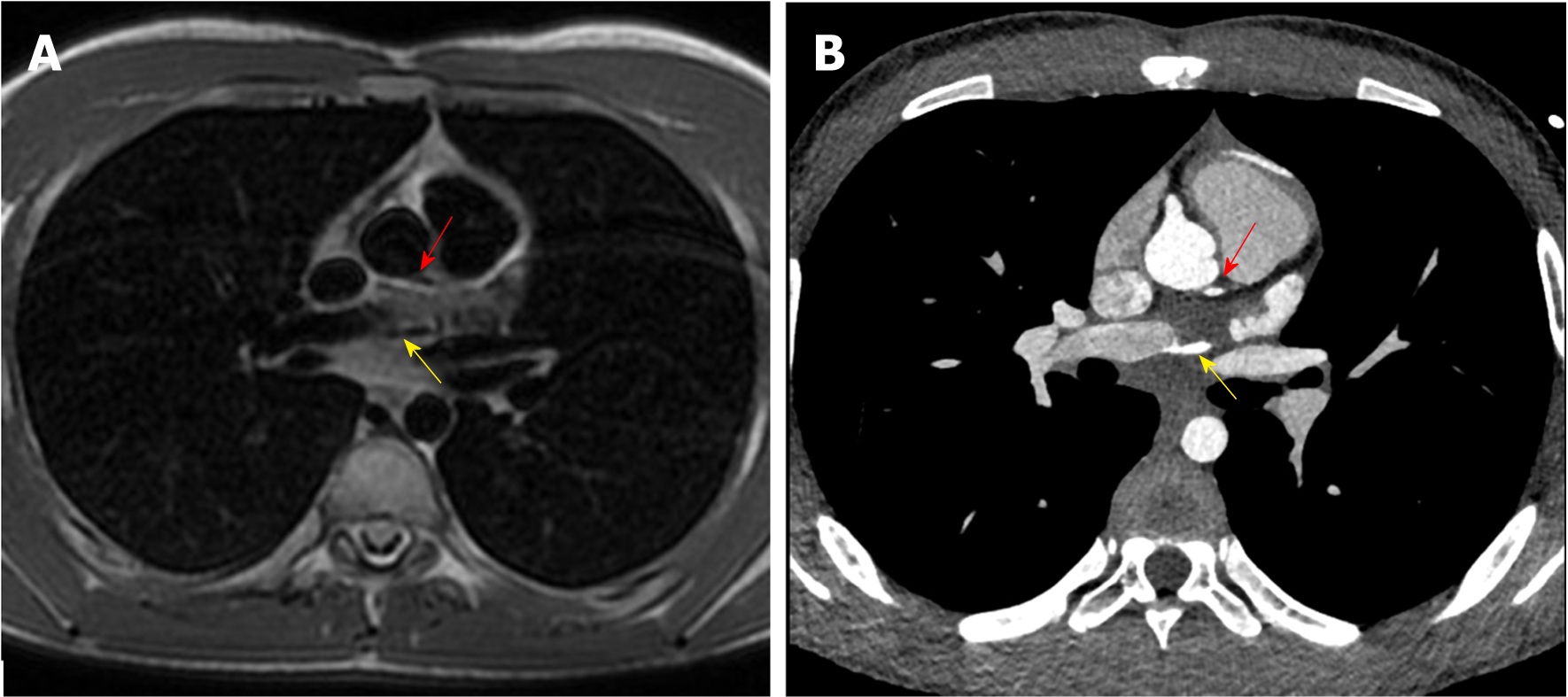
Figure 1 Origin of left circumflex artery and small diagonal branch.
A: Origin of left circumflex artery from right pulmonary artery (yellow arrow) and origin of a small diagonal branch from the left coronary sinus for the left ventricle (red arrow) evaluated with 1.5 T cardiac magnetic resonance; B: Origin of left circumflex artery from right pulmonary artery (yellow arrow) and origin of a small diagonal branch from the left coronary sinus for the left ventricle (red arrow) evaluated with third generation dual source cardiac computed tomography.
- Citation: Schicchi N, Fogante M, Giuseppetti GM, Giovagnoni A. Diagnostic detection with cardiac tomography and resonance of extremely rare coronary anomaly: A case report and review of literature. World J Clin Cases 2019; 7(5): 628-635
- URL: https://www.wjgnet.com/2307-8960/full/v7/i5/628.htm
- DOI: https://dx.doi.org/10.12998/wjcc.v7.i5.628


