Copyright
©The Author(s) 2019.
World J Clin Cases. Feb 26, 2019; 7(4): 525-531
Published online Feb 26, 2019. doi: 10.12998/wjcc.v7.i4.525
Published online Feb 26, 2019. doi: 10.12998/wjcc.v7.i4.525
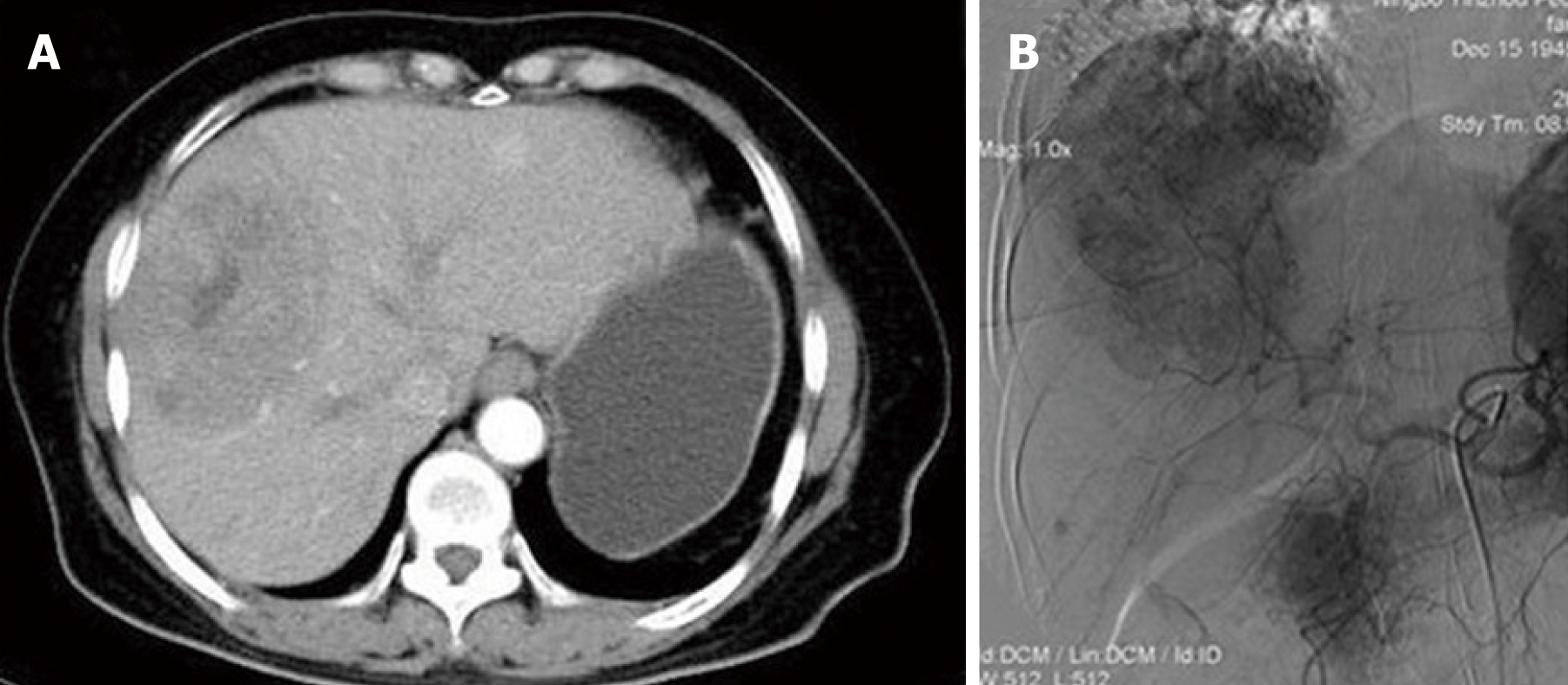
Figure 1 Imagining features before treatment.
A: Computed tomography showed a mass with inhomogeneous density, mild delayed enhancement and central necrosis; B: Hepatic angiography demonstrated a single mass with hypervascular tumor staining and dilated feeding arteries.
- Citation: Zhu KL, Cai XJ. Primary hepatic leiomyosarcoma successfully treated by transcatheter arterial chemoembolization: A case report. World J Clin Cases 2019; 7(4): 525-531
- URL: https://www.wjgnet.com/2307-8960/full/v7/i4/525.htm
- DOI: https://dx.doi.org/10.12998/wjcc.v7.i4.525


