Copyright
©The Author(s) 2019.
World J Clin Cases. Feb 26, 2019; 7(4): 508-515
Published online Feb 26, 2019. doi: 10.12998/wjcc.v7.i4.508
Published online Feb 26, 2019. doi: 10.12998/wjcc.v7.i4.508
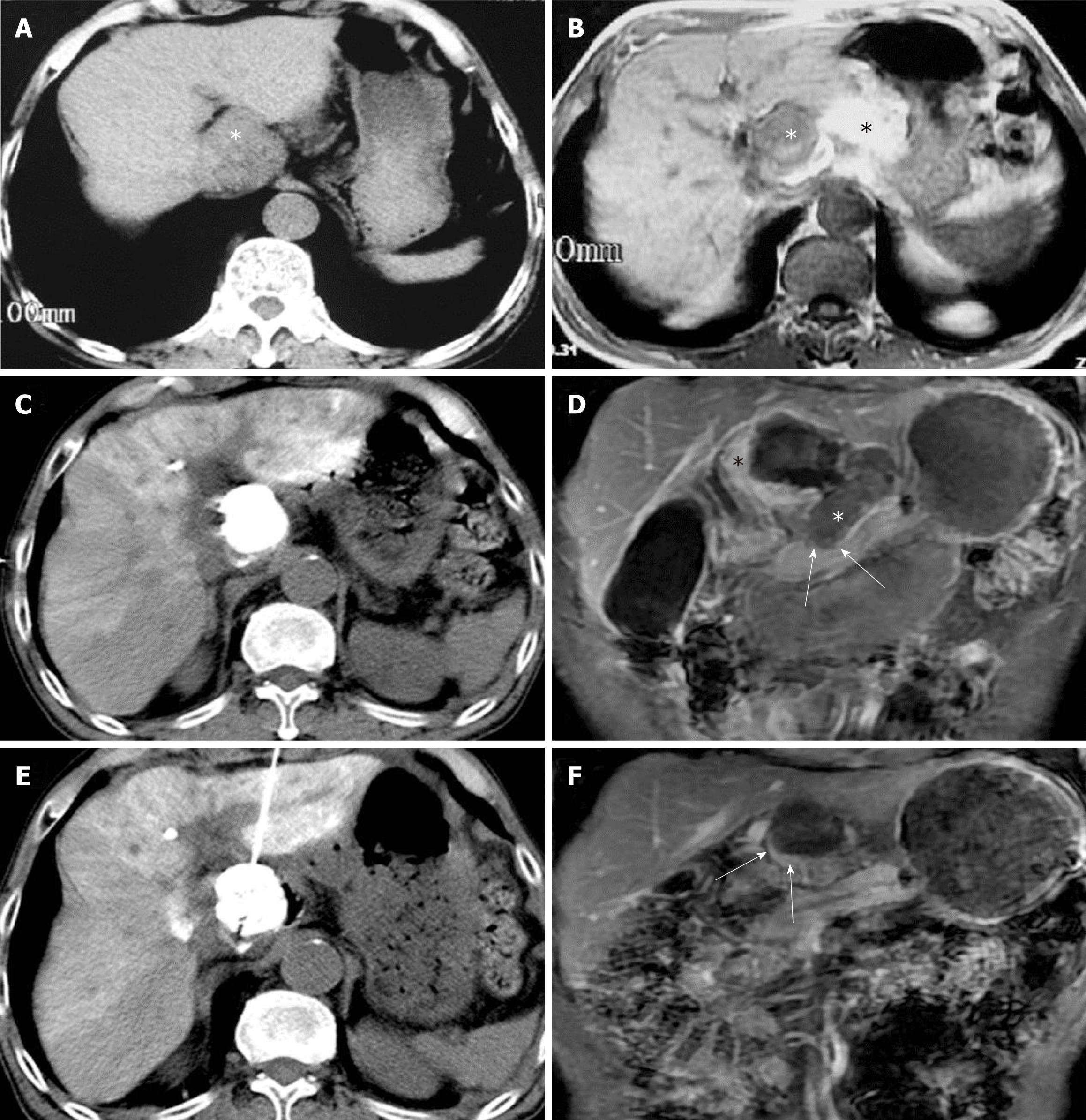
Figure 3 Preoperative computed tomography and magnetic resonance imaging images of a 73-year-old male showing a large intrahepatic tumor in the caudate lobe.
A and B: A large intrahepatic tumor in the caudate lobe was confirmed as hepatocellular carcinoma by histopathology. A hyperintense mass was noted in the pancreatic neck region on T1-weighted imaging (black asterisk, B). This was shown to be a hematoma on subsequent exploratory laparotomy; C: CT images showing that lipiodol deposition involved almost the entire area of the lesion in the caudate lobe after TACE; D: Peripheral residual tumor medial to the necrotic cavity was demonstrated in a coronal post-contrast T1-weighted image (black asterisk). The non-enhancing soft tissue in the porta hepatis region inferior to the intrahepatic lesion and adjacent to the neck of pancreas (white arrows) is consistent with the shrunken hematoma after exploratory laparotomy (white asterisk). E: CT image showing the needle in the tumor during HRFA; F: A thin enhancing rim-like tissue inferior to the necrotic cavity (white arrows) is evident on the post-contrast T1-weighted image after HRFA therap. The necrotic region does not show enhancement. No relapse was detected during follow-up MRI examinations. This enhancing rim probably represents reactive granulation tissues, rather than residual tumor.
- Citation: Deng HX, Huang JH, Lau WY, Ai F, Chen MS, Huang ZM, Zhang TQ, Zuo MX. Hydrochloric acid enhanced radiofrequency ablation for treatment of large hepatocellular carcinoma in the caudate lobe: Report of three cases. World J Clin Cases 2019; 7(4): 508-515
- URL: https://www.wjgnet.com/2307-8960/full/v7/i4/508.htm
- DOI: https://dx.doi.org/10.12998/wjcc.v7.i4.508


