Copyright
©The Author(s) 2019.
World J Clin Cases. Feb 26, 2019; 7(4): 508-515
Published online Feb 26, 2019. doi: 10.12998/wjcc.v7.i4.508
Published online Feb 26, 2019. doi: 10.12998/wjcc.v7.i4.508
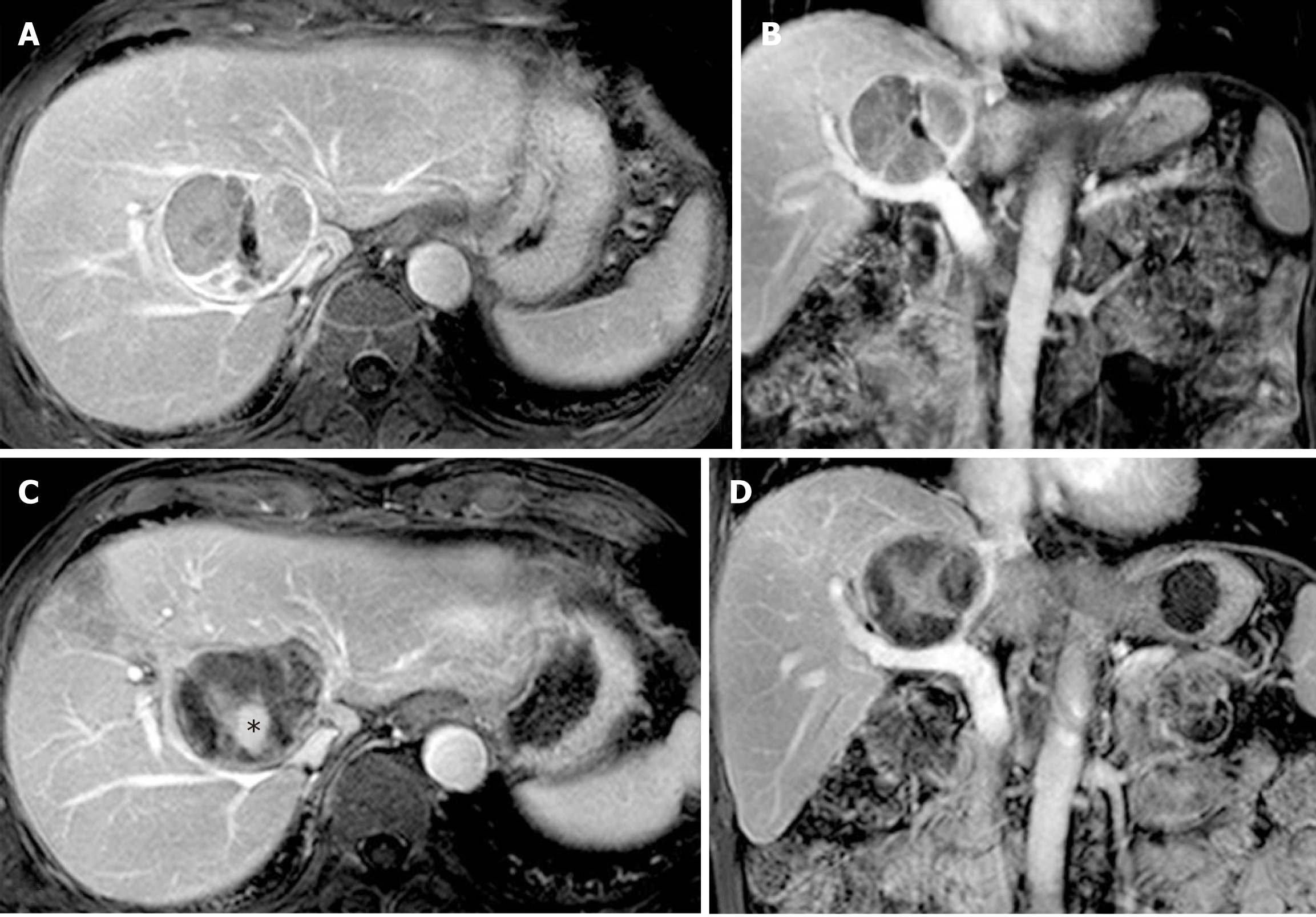
Figure 2 A well-defined active caudate lobe tumor adjacent to the right hepatic vein in a 69-year-old man with a history of tongue cancer after two sessions of TACE.
A and B: The enhanced part was predominant and had poor lipiodol deposition consistent with active tumor tissues; C and D: Post-contrast magnetic resonance imaging (MRI) images after HRFA therapy show a non-enhanced mass with hypointensity. The central irregular hyperintensity (black asterisk, C) was caused by the hemorrhagic content of the necrotic cavity, rather than by contrast enhancement. Relapse was not detected in the most recent MRI examination (data not shown).
- Citation: Deng HX, Huang JH, Lau WY, Ai F, Chen MS, Huang ZM, Zhang TQ, Zuo MX. Hydrochloric acid enhanced radiofrequency ablation for treatment of large hepatocellular carcinoma in the caudate lobe: Report of three cases. World J Clin Cases 2019; 7(4): 508-515
- URL: https://www.wjgnet.com/2307-8960/full/v7/i4/508.htm
- DOI: https://dx.doi.org/10.12998/wjcc.v7.i4.508


