Copyright
©The Author(s) 2019.
World J Clin Cases. Feb 26, 2019; 7(4): 508-515
Published online Feb 26, 2019. doi: 10.12998/wjcc.v7.i4.508
Published online Feb 26, 2019. doi: 10.12998/wjcc.v7.i4.508
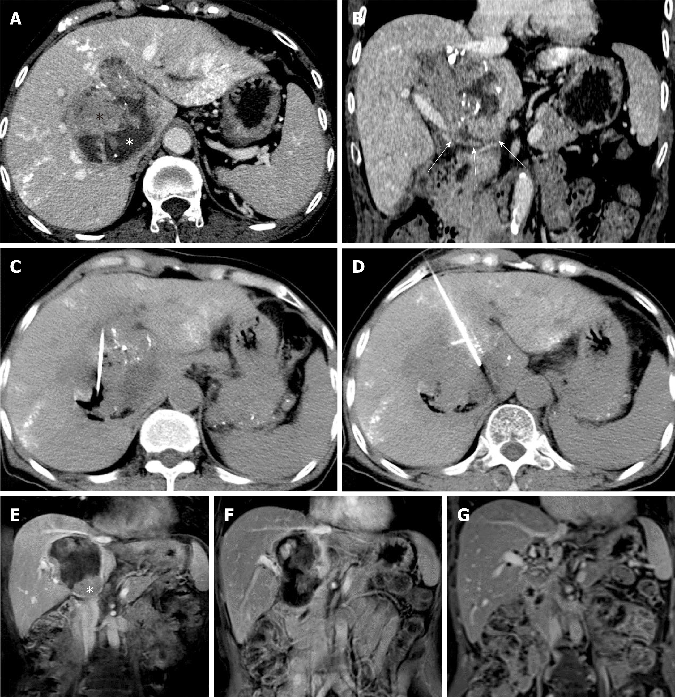
Figure 1 Arterial-phase computed tomography images showing a large caudate lobe tumor enhanced after transarterial chemoembolization in a 61-year-old woman with confirmed hepatocellular carcinoma.
A: The tumor with poor lipiodol deposition was predominantly composed by isodense tissue (black asterisk), representing the contrast enhanced active part of the tumor and the hypodense necrotic region without enhancement (white asterisk). The hyperdensity in the hepatic parenchyma represents lipiodol deposition after TACE; B: The caudate lobe and porta hepatis were involved (white arrows); C and D: Computed tomography images taken during the ablation show precise placement of the RF electrode into the active regions of the tumor, and the area of destruction can be seen easily; E: The coronal magnetic resonance image (MRI) shows an enhanced inferior residual tumor after the first session of HRFA (white asterisk); F: No active tissue was detected after the second session of HRFA; G: The latest coronal post-contrast T1-weighted image shows focal atrophy and fibrosis formation, resulting in an irregular, non-active region in the caudate lobe about 20 mm in diameter. The bile ducts were slightly dilated on the latest follow-up MRI.
- Citation: Deng HX, Huang JH, Lau WY, Ai F, Chen MS, Huang ZM, Zhang TQ, Zuo MX. Hydrochloric acid enhanced radiofrequency ablation for treatment of large hepatocellular carcinoma in the caudate lobe: Report of three cases. World J Clin Cases 2019; 7(4): 508-515
- URL: https://www.wjgnet.com/2307-8960/full/v7/i4/508.htm
- DOI: https://dx.doi.org/10.12998/wjcc.v7.i4.508


