Copyright
©The Author(s) 2019.
World J Clin Cases. Feb 26, 2019; 7(4): 482-488
Published online Feb 26, 2019. doi: 10.12998/wjcc.v7.i4.482
Published online Feb 26, 2019. doi: 10.12998/wjcc.v7.i4.482
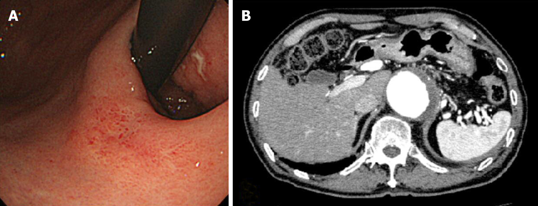
Figure 2 Objective image findings after chemotherapy.
A: EGD finding after chemotherapy. The lesion became unclear. It appeared smooth and protruded, and looked like a submucosal tumor; B: Abdominal CT with contrast after chemotherapy showed multiple lesions had disappeared and no evidence of disease in the liver. EGD: Esophagogastroduodenoscopy; CT: Computed tomography.
- Citation: Hayashi K, Suzuki S, Ikehara H, Okuno H, Irie A, Esaki M, Kusano C, Gotoda T, Moriyama M. Endoscopic resection for residual lesion of metastatic gastric cancer: A case report. World J Clin Cases 2019; 7(4): 482-488
- URL: https://www.wjgnet.com/2307-8960/full/v7/i4/482.htm
- DOI: https://dx.doi.org/10.12998/wjcc.v7.i4.482


