Copyright
©The Author(s) 2019.
World J Clin Cases. Feb 6, 2019; 7(3): 373-381
Published online Feb 6, 2019. doi: 10.12998/wjcc.v7.i3.373
Published online Feb 6, 2019. doi: 10.12998/wjcc.v7.i3.373
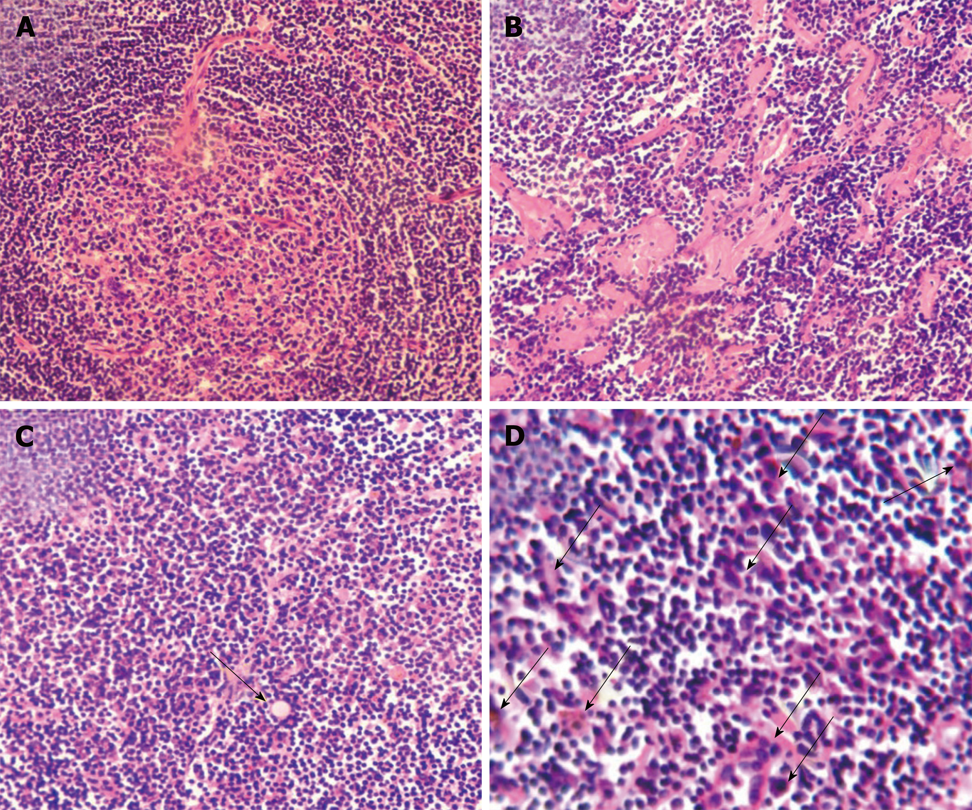
Figure 1 Major pathohistological features of mixed type Castleman disease.
Tissue sections (5 μm) were stained with hematoxylin and eosin. Histological characteristics in representative images include hyperplasia of follicular lymphoids concentrically layered around vascularized and degenerative germinal centers and shortage of lymphatic sinuses (A), hyaline degeneration (B), existence of Russell’s body (arrow) (C), and abundant proliferating plasma cells (arrows) (C and D). Magnification, × 200 (A-C) and × 400 (D).
- Citation: Zhai B, Ren HY, Li WD, Reddy S, Zhang SJ, Sun XY. Castleman disease presenting with jaundice: A case report and review of literature. World J Clin Cases 2019; 7(3): 373-381
- URL: https://www.wjgnet.com/2307-8960/full/v7/i3/373.htm
- DOI: https://dx.doi.org/10.12998/wjcc.v7.i3.373


