Copyright
©The Author(s) 2019.
World J Clin Cases. Dec 26, 2019; 7(24): 4398-4406
Published online Dec 26, 2019. doi: 10.12998/wjcc.v7.i24.4398
Published online Dec 26, 2019. doi: 10.12998/wjcc.v7.i24.4398
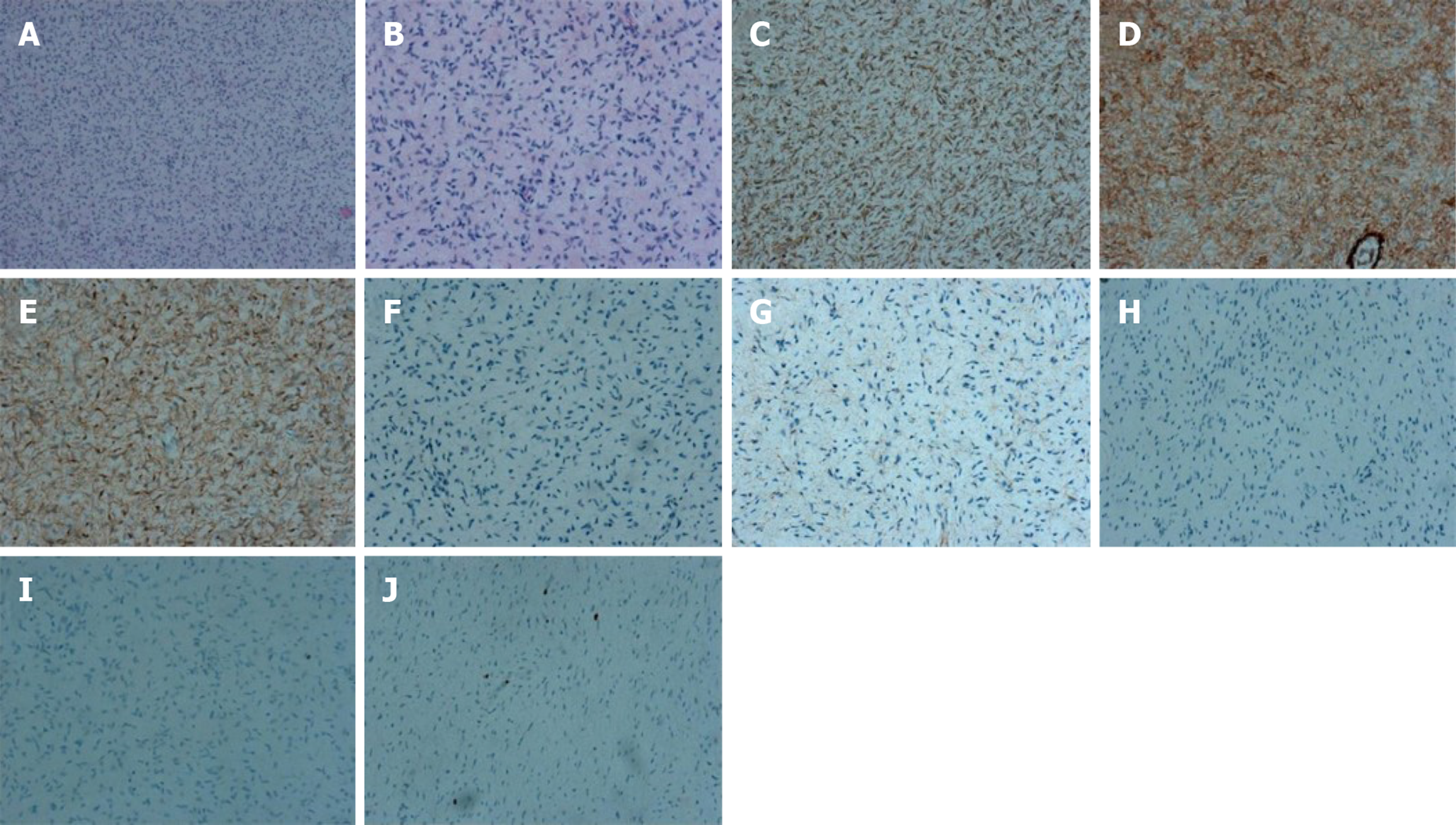
Figure 3 Microscopic and immunohistochemical features of multiple nodules from the skin.
A, B: Tumor composed of spindle cells with eosinophilic cytoplasm (A: × 100; B: × 200, hematoxylin-eosin staining); C: Positive for CD34 [3,3’-Diaminobenzidine (DAB) staining]; D: Positive for S-100 (DAB staining); E: Positive for VIM (DAB staining); F: Negative for CK (DAB staining); G: Negative for EMA (DAB staining); H: Negative for SMA (DAB staining); I: Negative for desmin (DAB staining); J: The percentage of Ki67 positive cells was approximately 2% (DAB staining).
- Citation: Kou YW, Zhang Y, Fu YP, Wang Z. KIT and platelet-derived growth factor receptor α wild-type gastrointestinal stromal tumor associated with neurofibromatosis type 1: Two case reports. World J Clin Cases 2019; 7(24): 4398-4406
- URL: https://www.wjgnet.com/2307-8960/full/v7/i24/4398.htm
- DOI: https://dx.doi.org/10.12998/wjcc.v7.i24.4398


