Copyright
©The Author(s) 2019.
World J Clin Cases. Dec 26, 2019; 7(24): 4307-4313
Published online Dec 26, 2019. doi: 10.12998/wjcc.v7.i24.4307
Published online Dec 26, 2019. doi: 10.12998/wjcc.v7.i24.4307
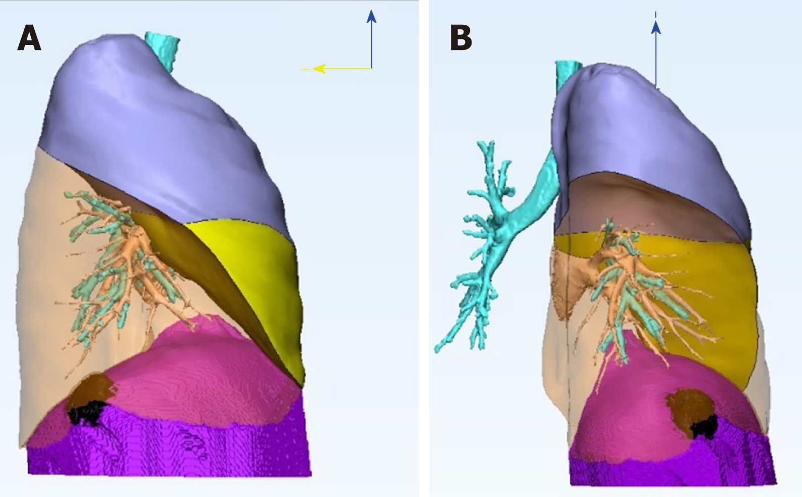
Figure 3 Three-dimension reconstruction image illustrating the relationship of the tumor and neighboring organs.
We mark the right uppeer lobe in lavender color, the right middle lobe in yellow, and the diaphrgam in purple. The right lower lobe is transparent to see the black lesion clearly. The three-dimension reconstruction image reveals a diaphragmatic lesion, without lung parenchyma involvement. A: Right lateral view of the right lung; B: Posterior view of the right lung.
- Citation: Chu PY, Lin KH, Kao HL, Peng YJ, Huang TW. Three-dimensional image simulation of primary diaphragmatic hemangioma: A case report. World J Clin Cases 2019; 7(24): 4307-4313
- URL: https://www.wjgnet.com/2307-8960/full/v7/i24/4307.htm
- DOI: https://dx.doi.org/10.12998/wjcc.v7.i24.4307


