Copyright
©The Author(s) 2019.
World J Clin Cases. Dec 26, 2019; 7(24): 4307-4313
Published online Dec 26, 2019. doi: 10.12998/wjcc.v7.i24.4307
Published online Dec 26, 2019. doi: 10.12998/wjcc.v7.i24.4307
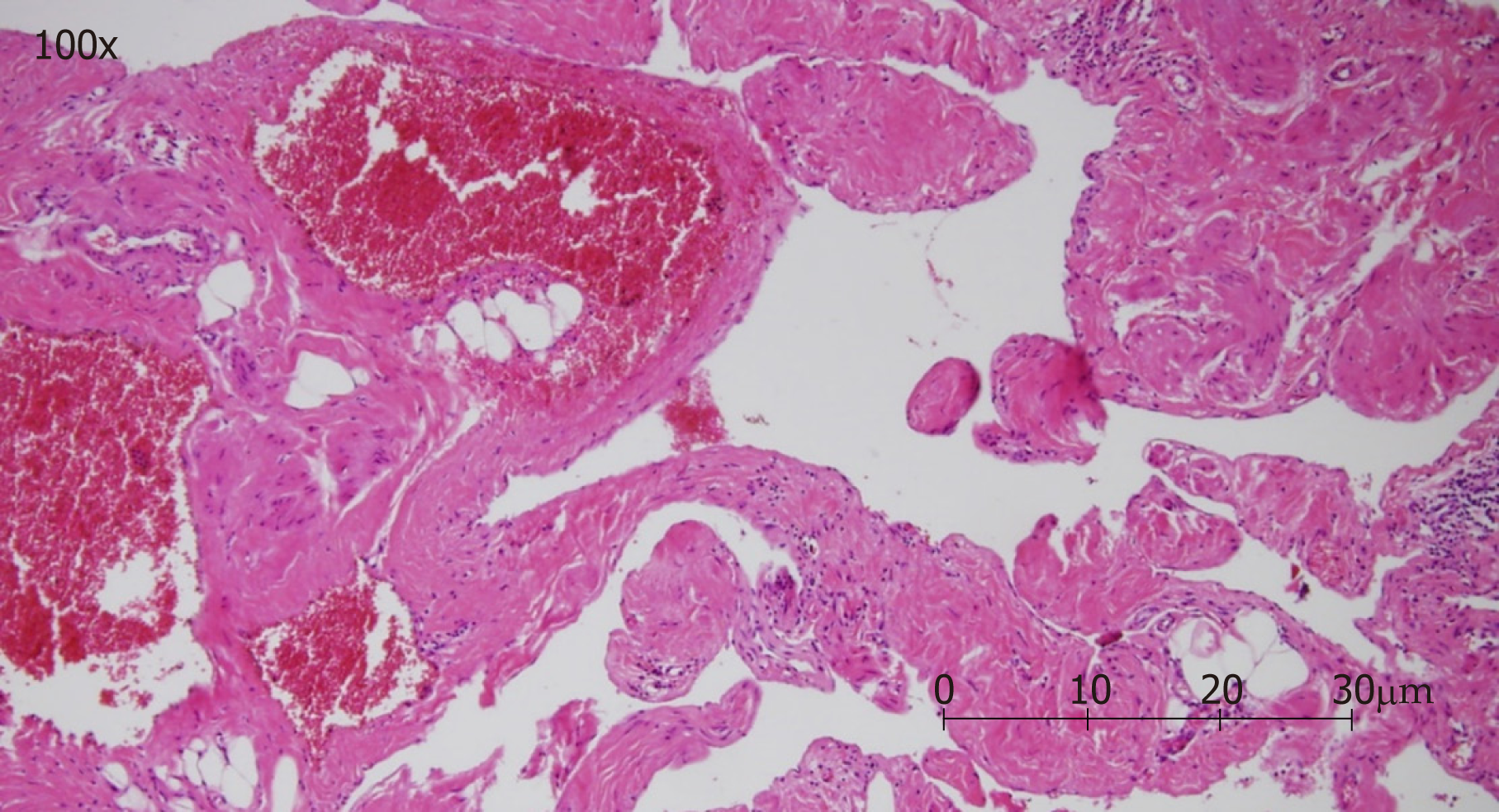
Figure 2 Pathologic image showing a vascular lesion composed of dilated cavernous vascular spaces separated by irregular vascular walls microscopically.
The pathologic findings (hematoxylin and eosin staining; magnification, 100×) are compatible with those of cavernous hemangioma.
- Citation: Chu PY, Lin KH, Kao HL, Peng YJ, Huang TW. Three-dimensional image simulation of primary diaphragmatic hemangioma: A case report. World J Clin Cases 2019; 7(24): 4307-4313
- URL: https://www.wjgnet.com/2307-8960/full/v7/i24/4307.htm
- DOI: https://dx.doi.org/10.12998/wjcc.v7.i24.4307


