Copyright
©The Author(s) 2019.
World J Clin Cases. Dec 26, 2019; 7(24): 4307-4313
Published online Dec 26, 2019. doi: 10.12998/wjcc.v7.i24.4307
Published online Dec 26, 2019. doi: 10.12998/wjcc.v7.i24.4307
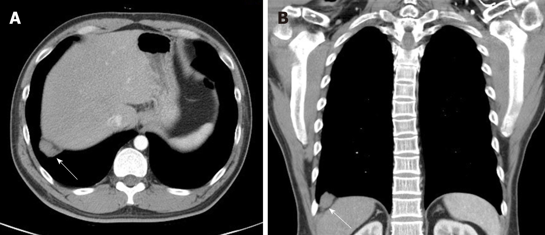
Figure 1 Contrast-enhanced chest computed tomography images illustrating a poorly-enhanced lesion (arrow) in the right basal lung, abutting the right diaphragm.
A: Axial view; B: Coronal view.
- Citation: Chu PY, Lin KH, Kao HL, Peng YJ, Huang TW. Three-dimensional image simulation of primary diaphragmatic hemangioma: A case report. World J Clin Cases 2019; 7(24): 4307-4313
- URL: https://www.wjgnet.com/2307-8960/full/v7/i24/4307.htm
- DOI: https://dx.doi.org/10.12998/wjcc.v7.i24.4307


