Copyright
©The Author(s) 2019.
World J Clin Cases. Dec 26, 2019; 7(24): 4299-4306
Published online Dec 26, 2019. doi: 10.12998/wjcc.v7.i24.4299
Published online Dec 26, 2019. doi: 10.12998/wjcc.v7.i24.4299
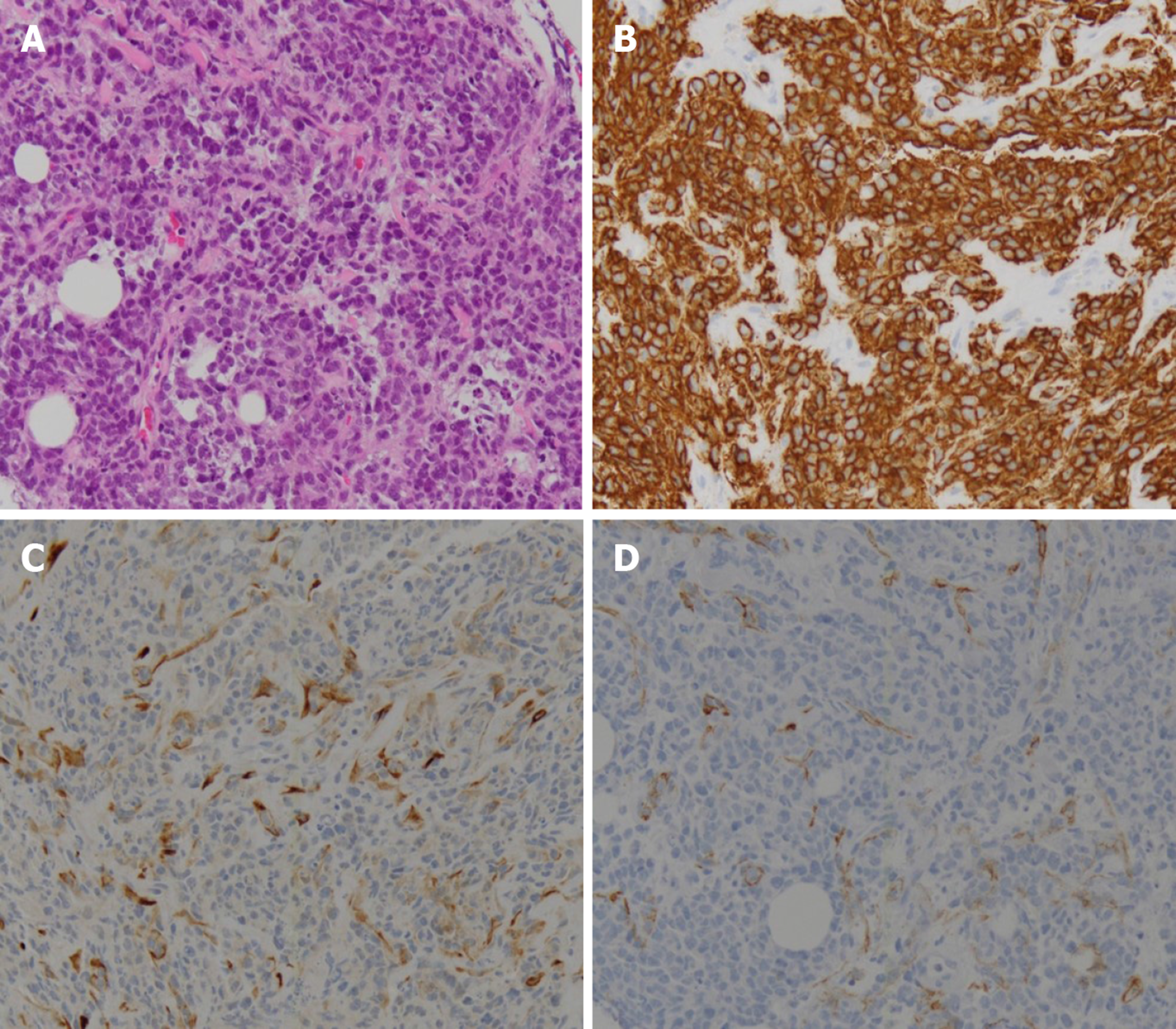
Figure 3 Pathologic findings.
Highly pleomorphic, atypical, small, round, blue cells were observed infiltrating the fibrocollagenous tissue (A). On immunohistochemical testing, these cells were positive for CD20 (B) and LCA, but they were negative for the mesothelial cell marker D2-40 (C) and the epithelial cell marker cytokeratin (D).
- Citation: Kim HB, Hong R, Na YS, Choi WY, Park SG, Lee HJ. Isolated peritoneal lymphomatosis defined as post-transplant lymphoproliferative disorder after a liver transplant: A case report. World J Clin Cases 2019; 7(24): 4299-4306
- URL: https://www.wjgnet.com/2307-8960/full/v7/i24/4299.htm
- DOI: https://dx.doi.org/10.12998/wjcc.v7.i24.4299


