Copyright
©The Author(s) 2019.
World J Clin Cases. Dec 6, 2019; 7(23): 4119-4129
Published online Dec 6, 2019. doi: 10.12998/wjcc.v7.i23.4119
Published online Dec 6, 2019. doi: 10.12998/wjcc.v7.i23.4119
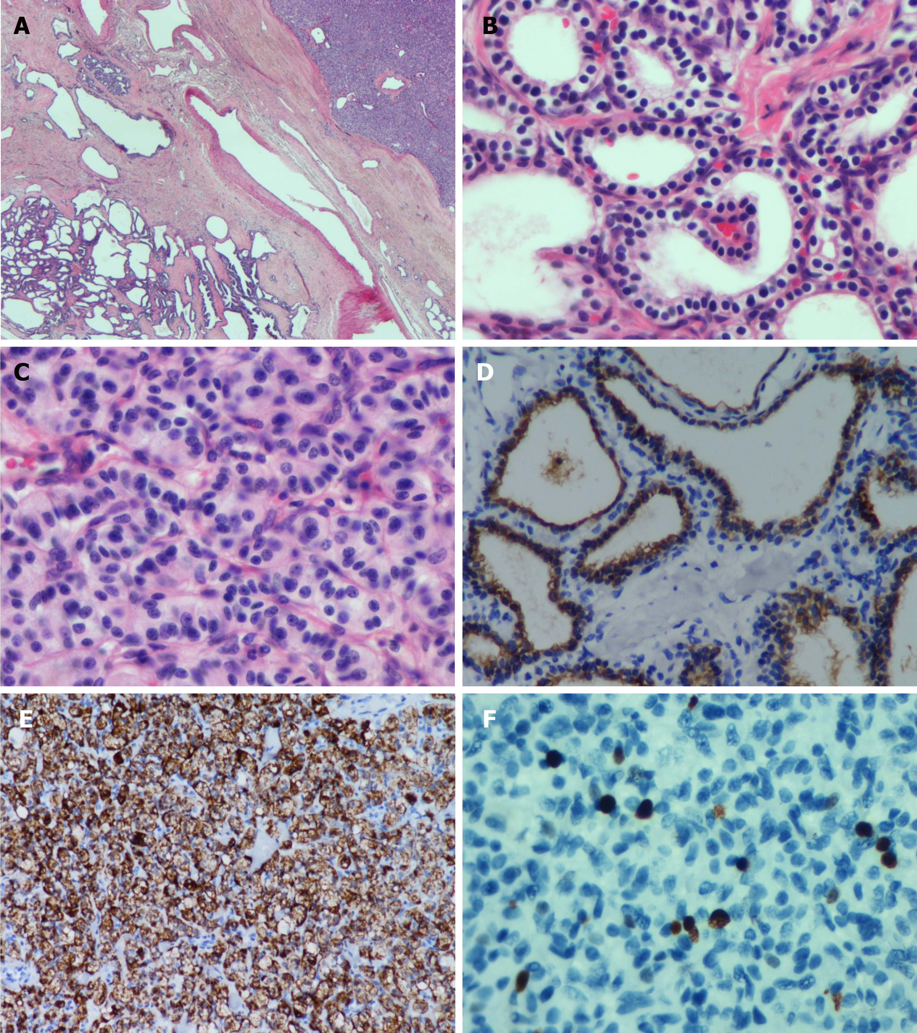
Figure 3 Histological morphology of the diffuse cystic lesions and the solid mass appears significantly different.
A: The two components of the pancreatic tumor were separated by compressed fibrous tissue without any overlapping zone (Hematoxylin and eosin staining; original magnification, 20×); B: The diffuse cystic area was composed of prominent microcystic serous cystadenomas lined with a single layer of cubic to flattened epithelium with clear cytoplasm; the nuclei were uniformly small, round-to-oval, and bland with dense homogenous chromatin (Hematoxylin and eosin staining; original magnification, 400×); C: The solid tumor in the head of the pancreas demonstrated a well-differentiated grade 2 pancreatic neuroendocrine tumor (PanNET). The tumor cells were arranged in a ribbon-like and trabecular architecture with a fine chromatin and abundant microvasculature (Hematoxylin and eosin staining; original magnification, 400×); D: Immunohistochemically, the lining epithelium of the diffuse serous cystadenoma stained positive for cytokeratin7 (EnVision two-step method; original magnification, 400×); E: The low-grade PanNET was positive for chromogranin A (EnVision two-step method; original magnification, 200×); F: The Ki67 labeling index of the PanNET was 5% (EnVision two-step method; original magnification, 400×).
- Citation: Xu YM, Li ZW, Wu HY, Fan XS, Sun Q. Mixed serous-neuroendocrine neoplasm of the pancreas: A case report and review of the literature. World J Clin Cases 2019; 7(23): 4119-4129
- URL: https://www.wjgnet.com/2307-8960/full/v7/i23/4119.htm
- DOI: https://dx.doi.org/10.12998/wjcc.v7.i23.4119


