Copyright
©The Author(s) 2019.
World J Clin Cases. Dec 6, 2019; 7(23): 4119-4129
Published online Dec 6, 2019. doi: 10.12998/wjcc.v7.i23.4119
Published online Dec 6, 2019. doi: 10.12998/wjcc.v7.i23.4119
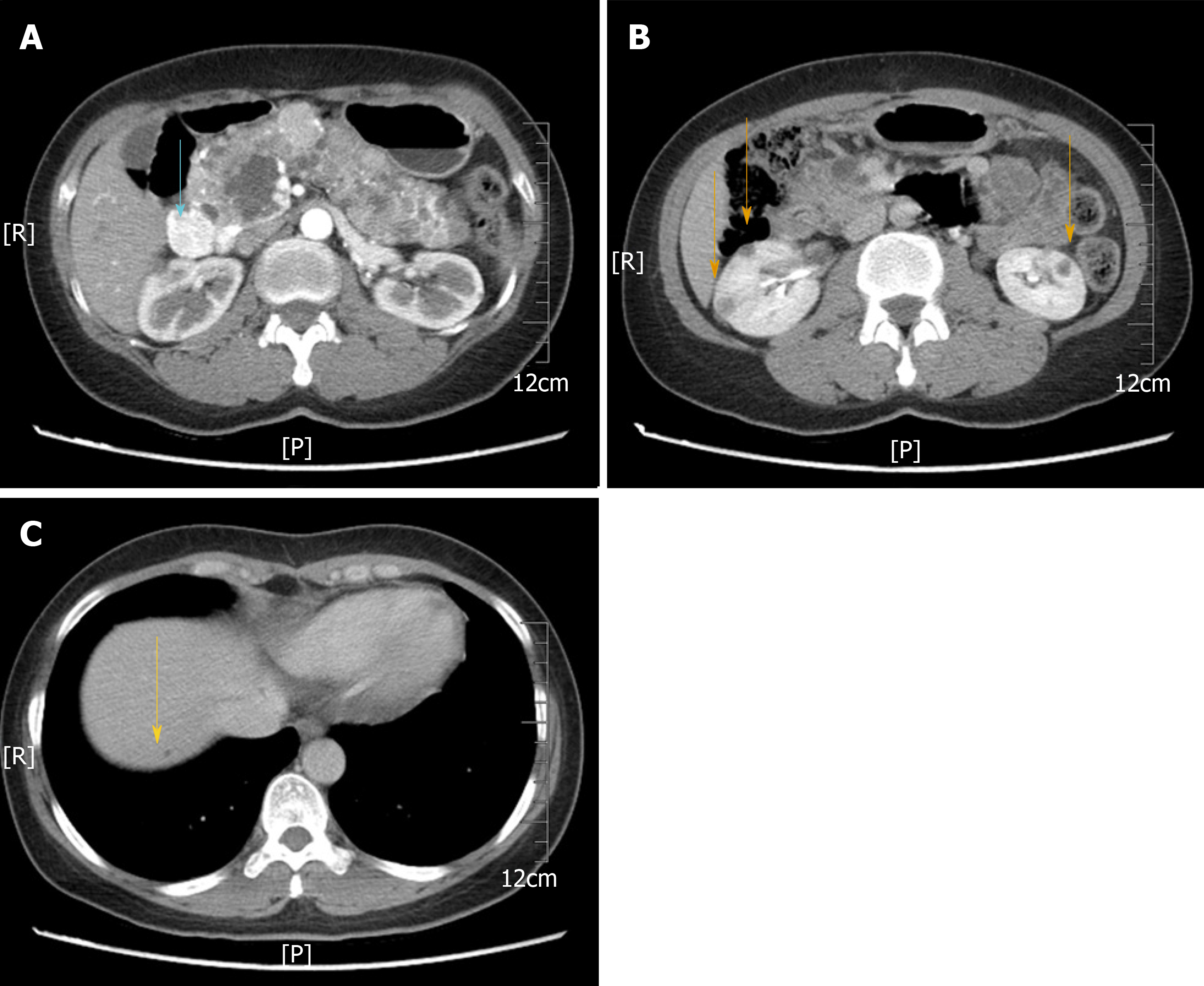
Figure 1 Abdominal computed tomography images.
A: Computed tomography image showing multiple cystic lesions involving the whole pancreas ranging in diameter from 0.4 to 2 cm. An enhanced mass was also revealed, 2.2 cm in diameter, in the head of the pancreas (blue arrow); B: Multiple cysts were found in the kidneys bilaterally (orange arrow); C: A small cyst in the right lobe of the liver (yellow arrow).
- Citation: Xu YM, Li ZW, Wu HY, Fan XS, Sun Q. Mixed serous-neuroendocrine neoplasm of the pancreas: A case report and review of the literature. World J Clin Cases 2019; 7(23): 4119-4129
- URL: https://www.wjgnet.com/2307-8960/full/v7/i23/4119.htm
- DOI: https://dx.doi.org/10.12998/wjcc.v7.i23.4119


