Copyright
©The Author(s) 2019.
World J Clin Cases. Dec 6, 2019; 7(23): 4111-4118
Published online Dec 6, 2019. doi: 10.12998/wjcc.v7.i23.4111
Published online Dec 6, 2019. doi: 10.12998/wjcc.v7.i23.4111
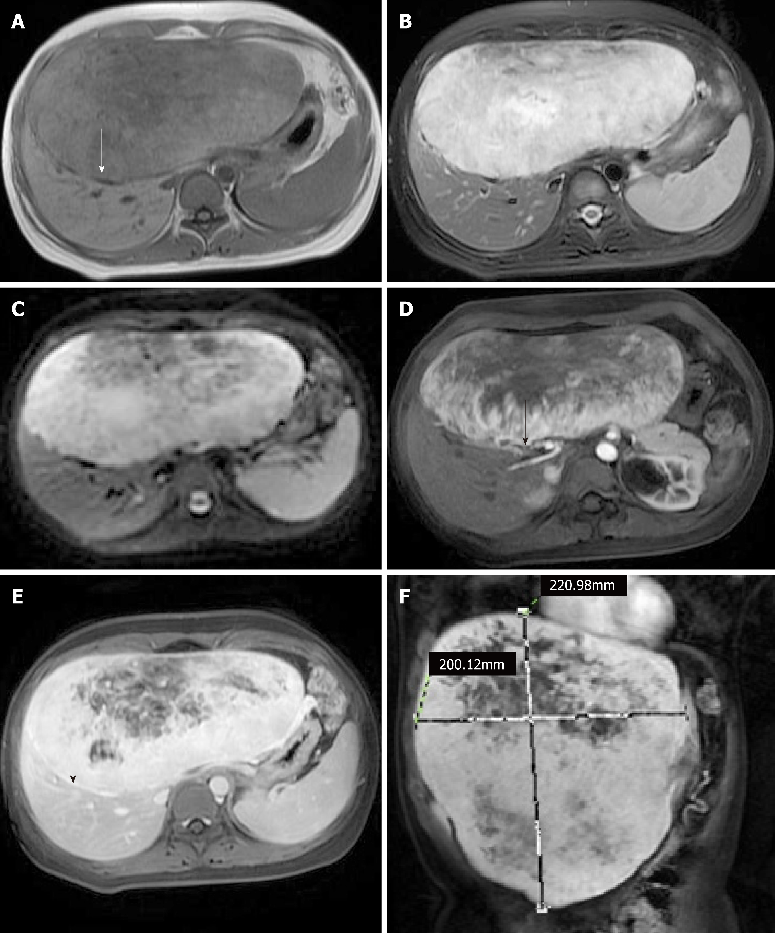
Figure 2 Hepatocellular adenoma on magnetic resonance imaging scans.
A: T1-weighted image. The fibrous capsule of the tumor was clearly visible (arrow); B: T2-weighted image; C: Diffusion-weighted imaging; D: Arterial phase of contrast-enhanced scan. Feeding arteries were visible on the verge of the tumor (arrow); E: Delayed phase of contrast-enhanced scan. The fibrous capsule of the tumor was markedly enhanced (arrow); F: Multiplanar reconstruction.
- Citation: Zheng LP, Hu CD, Wang J, Chen XJ, Shen YY. Radiological aspects of giant hepatocellular adenoma of the left liver: A case report. World J Clin Cases 2019; 7(23): 4111-4118
- URL: https://www.wjgnet.com/2307-8960/full/v7/i23/4111.htm
- DOI: https://dx.doi.org/10.12998/wjcc.v7.i23.4111


