Copyright
©The Author(s) 2019.
World J Clin Cases. Dec 6, 2019; 7(23): 4063-4074
Published online Dec 6, 2019. doi: 10.12998/wjcc.v7.i23.4063
Published online Dec 6, 2019. doi: 10.12998/wjcc.v7.i23.4063
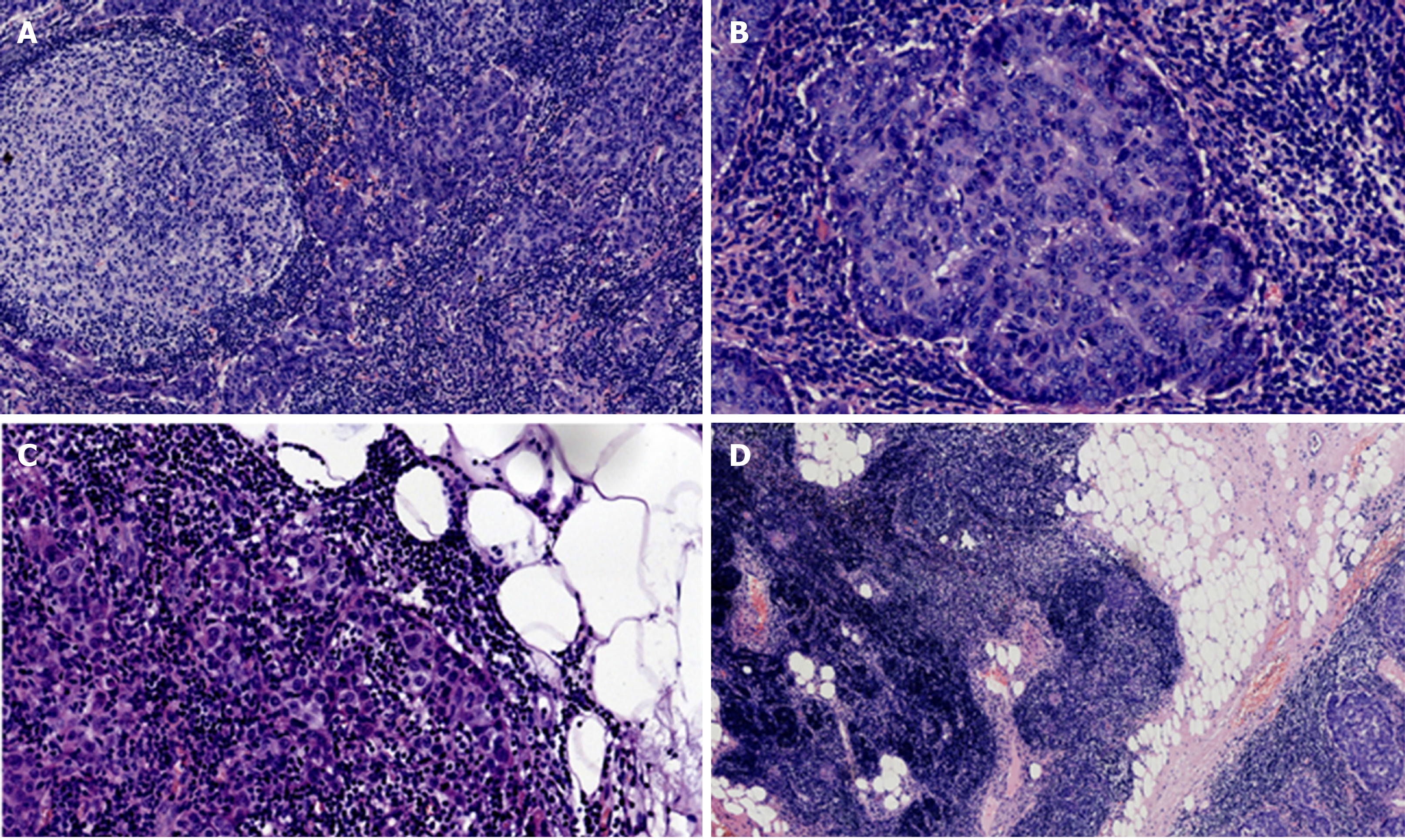
Figure 1 Hematoxylin and eosin staining of the tumor tissue.
A: Staining of lymphoid follicle formation sites in the stroma (×10); B: Image of an enlarged interstitial lymphoid follicle formation site at high magnification. The mitotic figures were 1-9/10 high power fields (×20); C: Staining of the fatty portion of the perimeter of the neoplasm. The white tissue is adipose tissue (×10); D: Histogram of the junction between the tumor and normal thymus tissues (×4).
- Citation: Wang B, Li K, Song QK, Wang XH, Yang L, Zhang HL, Zhong DR. Micronodular thymic tumor with lymphoid stroma: A case report and review of the literature. World J Clin Cases 2019; 7(23): 4063-4074
- URL: https://www.wjgnet.com/2307-8960/full/v7/i23/4063.htm
- DOI: https://dx.doi.org/10.12998/wjcc.v7.i23.4063


