Copyright
©The Author(s) 2019.
World J Clin Cases. Dec 6, 2019; 7(23): 4044-4051
Published online Dec 6, 2019. doi: 10.12998/wjcc.v7.i23.4044
Published online Dec 6, 2019. doi: 10.12998/wjcc.v7.i23.4044
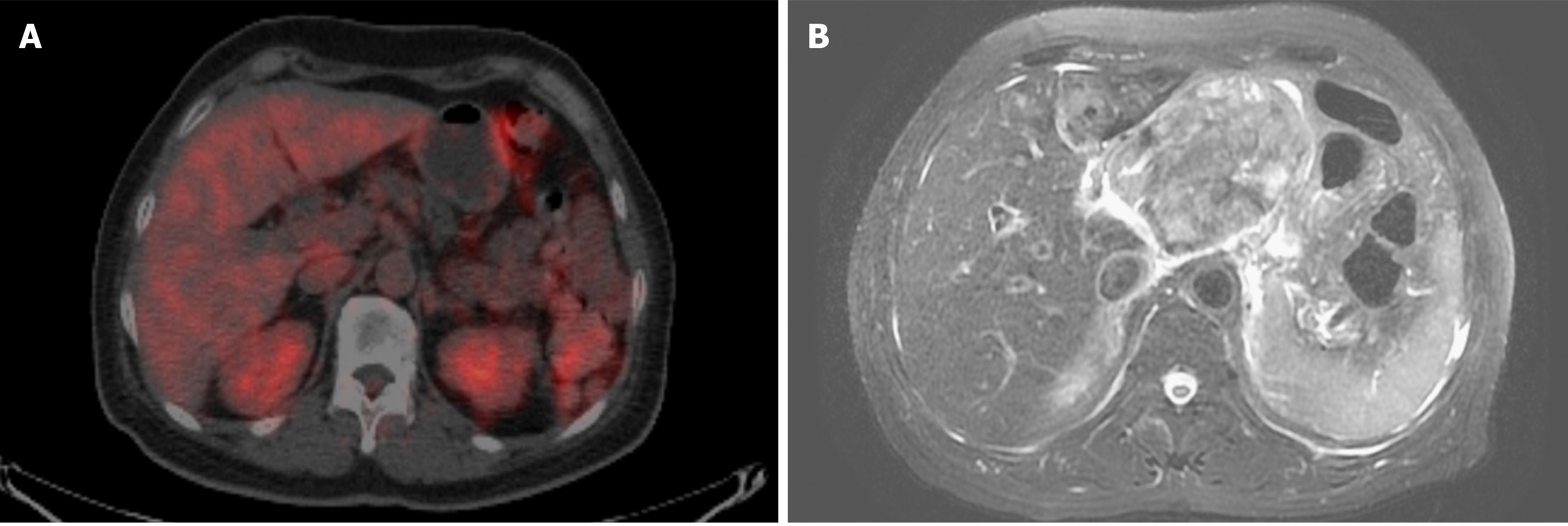
Figure 4 Images from abdominal positron emission tomography-computed tomography scans and magnetic resonance imaging.
A: positron emission tomography-computed tomography scan at initial presentation revealed no definite Fluorodeoxyglucose-avid tumor in the abdomen; B: Magnetic resonance imaging of the abdomen at 1.5 years after enucleation revealed a large mass at the upper abdomen abutting the pancreatic neck and body with a maximal diameter of at least 9.5 cm.
- Citation: Wang TW, Liu HW, Bee YS. Distant metastasis in choroidal melanoma with spontaneous corneal perforation and intratumoral calcification: A case report. World J Clin Cases 2019; 7(23): 4044-4051
- URL: https://www.wjgnet.com/2307-8960/full/v7/i23/4044.htm
- DOI: https://dx.doi.org/10.12998/wjcc.v7.i23.4044


