Copyright
©The Author(s) 2019.
World J Clin Cases. Dec 6, 2019; 7(23): 4044-4051
Published online Dec 6, 2019. doi: 10.12998/wjcc.v7.i23.4044
Published online Dec 6, 2019. doi: 10.12998/wjcc.v7.i23.4044
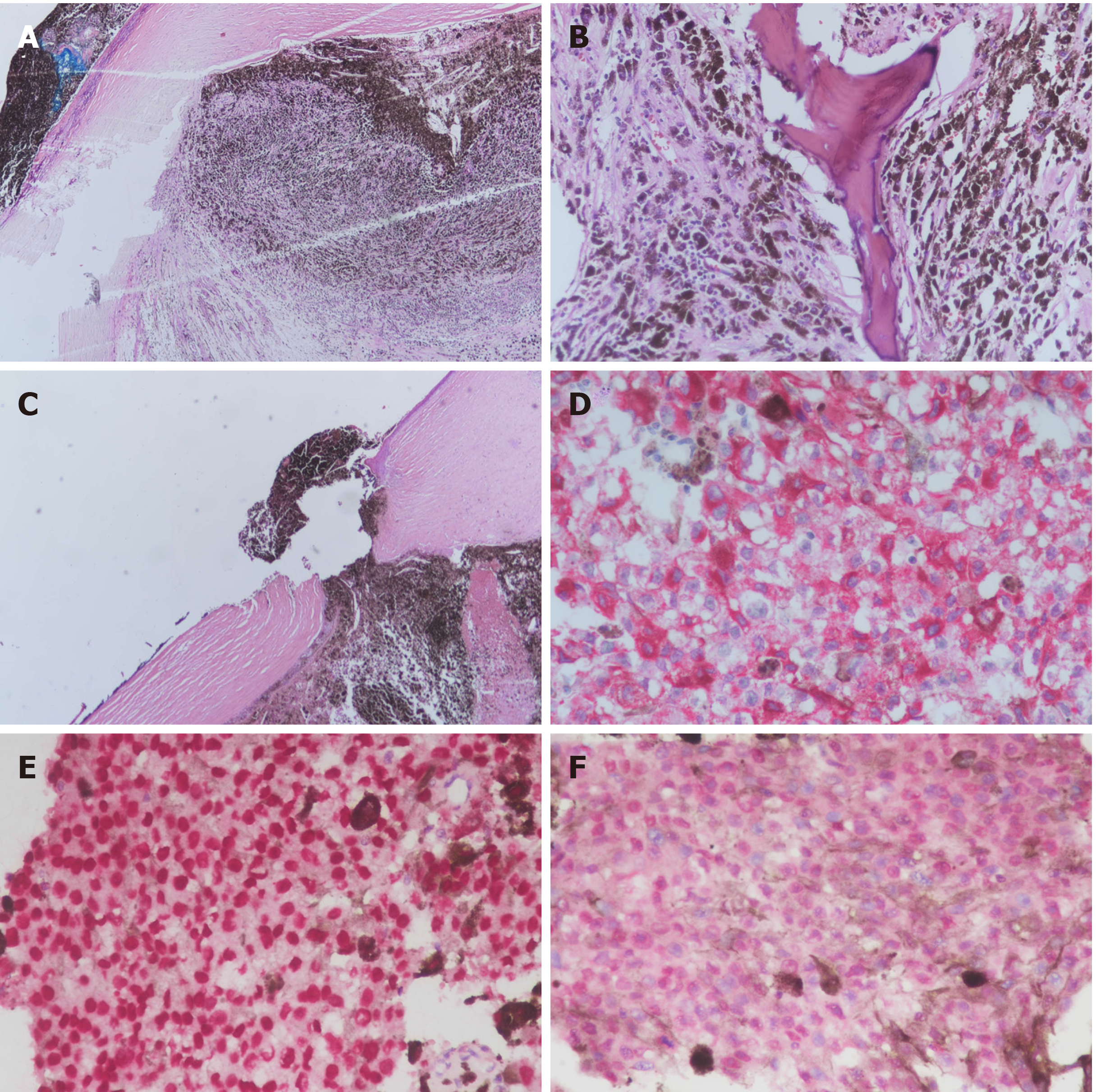
Figure 3 Histological findings of the resected tumor specimens.
A: Tumor cells occupied the entire eyeball (×40); B: Ossification was noted in the tumor (×200); C: Uveal tissue was confined in the region of the perforated cornea. Intact cornea layers and no corneal thinning or corneal ulcer was noted (×40); D: Immunohistochemistry (IHC) revealed diffusely positive melanin-A immunostaining in the cytoplasm of primary tumor cells (Red chromogen, ×400); E: IHC revealed diffusely positive SOX-10 immunostaining in the nuclei of primary tumor cells (Red chromogen, ×400); F: Primary epithelioid tumor cells showed strong nuclear labeling for BAP1 expression (Red chromogen, ×400).
- Citation: Wang TW, Liu HW, Bee YS. Distant metastasis in choroidal melanoma with spontaneous corneal perforation and intratumoral calcification: A case report. World J Clin Cases 2019; 7(23): 4044-4051
- URL: https://www.wjgnet.com/2307-8960/full/v7/i23/4044.htm
- DOI: https://dx.doi.org/10.12998/wjcc.v7.i23.4044


