Copyright
©The Author(s) 2019.
World J Clin Cases. Dec 6, 2019; 7(23): 4020-4028
Published online Dec 6, 2019. doi: 10.12998/wjcc.v7.i23.4020
Published online Dec 6, 2019. doi: 10.12998/wjcc.v7.i23.4020
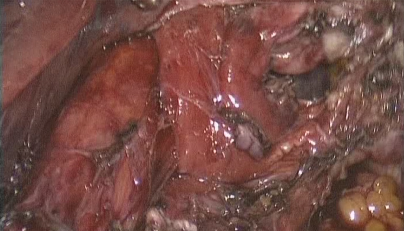
Figure 5 Final intraoperative aspect after dissection.
The esophagus can be seen at the left of the image, the hiatus is slightly enlarged, the diaphragmatic defect is located lateral to the esophageal hiatus and is separated by the left crus which is anatomically intact.
- Citation: Preda SD, Pătraşcu Ș, Ungureanu BS, Cristian D, Bințințan V, Nica CM, Calu V, Strâmbu V, Sapalidis K, Șurlin VM. Primary parahiatal hernias: A case report and review of the literature. World J Clin Cases 2019; 7(23): 4020-4028
- URL: https://www.wjgnet.com/2307-8960/full/v7/i23/4020.htm
- DOI: https://dx.doi.org/10.12998/wjcc.v7.i23.4020


