Copyright
©The Author(s) 2019.
World J Clin Cases. Nov 26, 2019; 7(22): 3895-3903
Published online Nov 26, 2019. doi: 10.12998/wjcc.v7.i22.3895
Published online Nov 26, 2019. doi: 10.12998/wjcc.v7.i22.3895
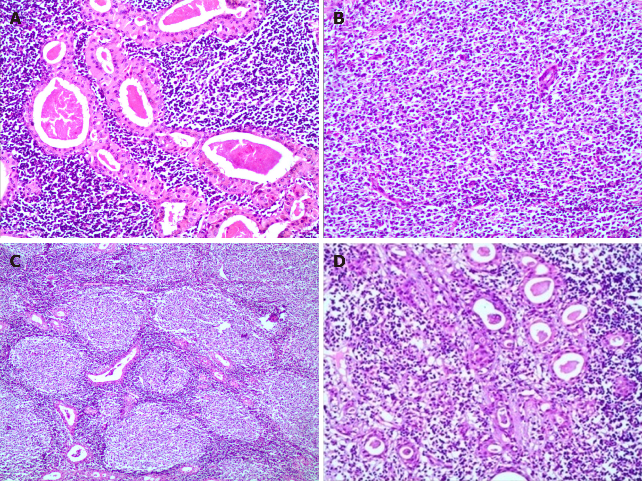
Figure 1 Morphological characteristic of Warthin’s tumor and lymphoma.
A: The bilayered oxyphilic, cuboidal or polygonal epithelium cells and lymphoid intraparernchymal components were observed; B, C: A lot of medium- to large- size lymphoid cells was observed diffusely in part of neoplasm (B) and a few secondary lymphoid follicles were seen at the centre or edge of neoplasm (C); D: At the border of epithelium, the lymphoepithelial lesions were identified. (hematoxylin-eosin staining, Magnification ×200)
- Citation: Wang CS, Chu X, Yang D, Ren L, Meng NL, Lv XX, Yun T, Cao YS. Diffuse large B-cell lymphoma arising from follicular lymphoma with warthin’s tumor of the parotid gland - immunophenotypic and genetic features: A case report. World J Clin Cases 2019; 7(22): 3895-3903
- URL: https://www.wjgnet.com/2307-8960/full/v7/i22/3895.htm
- DOI: https://dx.doi.org/10.12998/wjcc.v7.i22.3895


