Copyright
©The Author(s) 2019.
World J Clin Cases. Nov 26, 2019; 7(22): 3844-3850
Published online Nov 26, 2019. doi: 10.12998/wjcc.v7.i22.3844
Published online Nov 26, 2019. doi: 10.12998/wjcc.v7.i22.3844
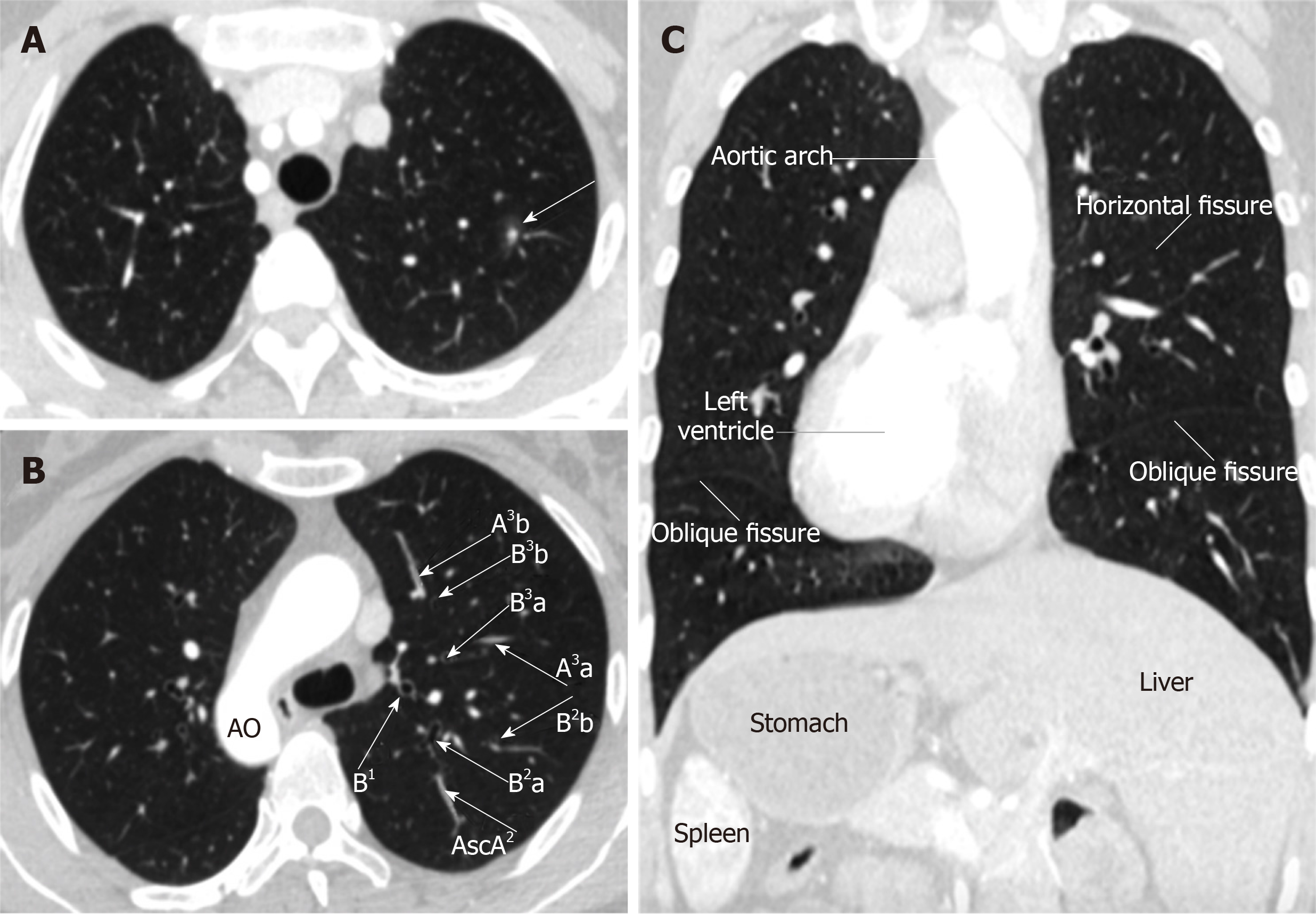
Figure 1 Computed tomography images.
A: Mixed ground-glass opacity measuring 1.2 cm in diameter with a solid component of approximately 0.6 cm in the posterior segment of the left upper lobe (LS2); B: Complete mirror image of the segmental vessels and bronchus of LS2; C: Situs inversus totalis (dextrocardia, aortic arch, spleen, and liver). AscA: Ascending artery.
- Citation: Wu YJ, Bao Y, Wang YL. Thoracoscopic segmentectomy assisted by three-dimensional computed tomography bronchography and angiography for lung cancer in a patient living with situs inversus totalis: A case report. World J Clin Cases 2019; 7(22): 3844-3850
- URL: https://www.wjgnet.com/2307-8960/full/v7/i22/3844.htm
- DOI: https://dx.doi.org/10.12998/wjcc.v7.i22.3844


