Copyright
©The Author(s) 2019.
World J Clin Cases. Nov 26, 2019; 7(22): 3832-3837
Published online Nov 26, 2019. doi: 10.12998/wjcc.v7.i22.3832
Published online Nov 26, 2019. doi: 10.12998/wjcc.v7.i22.3832
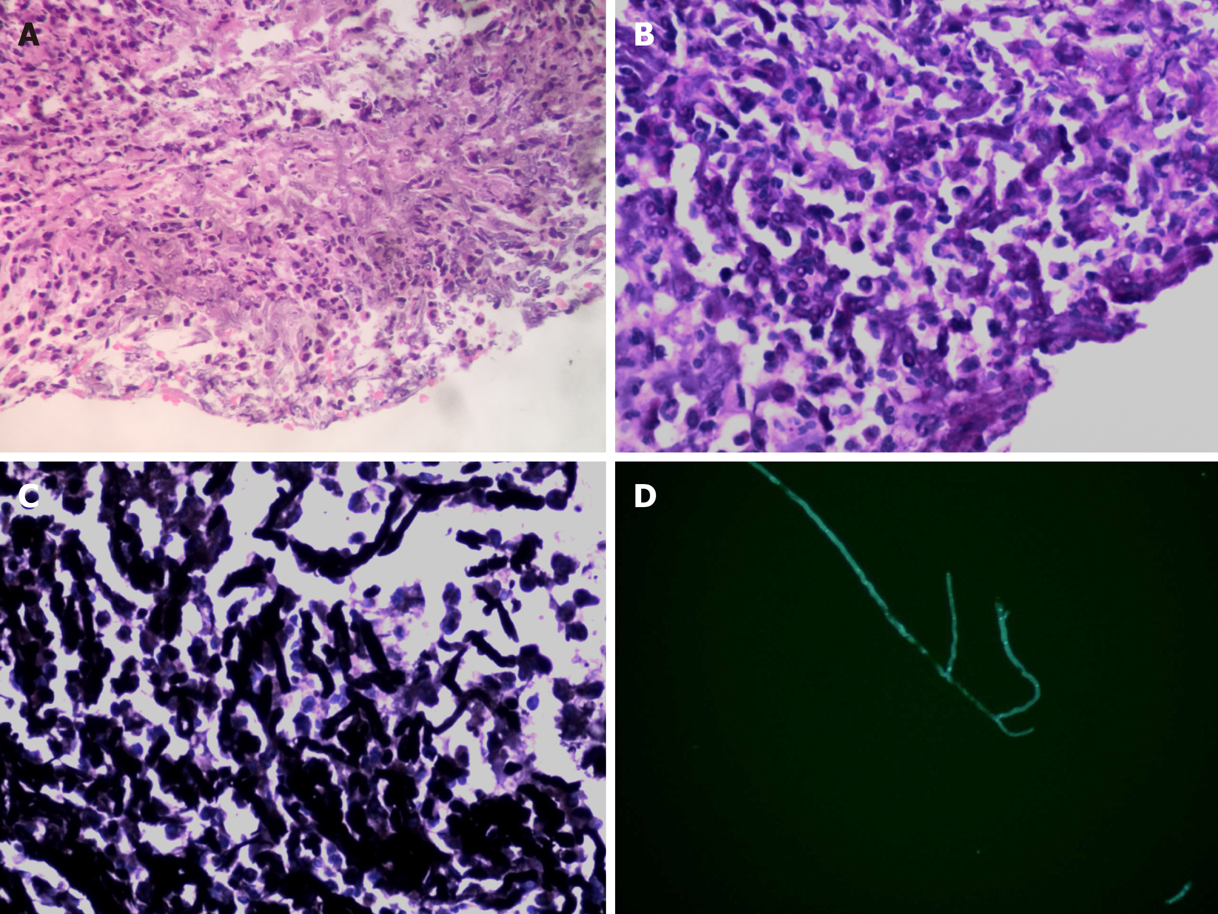
Figure 3 Histological and microbiological evidence of fungal infection.
A: Fungal elements showing the 45° branching hyphae within biopsies under bronchoscopy (hematoxylin–eosin stain, 400×); B: Periodic Acid-Schiff staining was positive (400×); C: Grocott staining was positive (400×); D: Fungal fluorescence staining of bronchial membrane brushing sample done on 2019-3-26 revealed branching septate hyphae.
- Citation: Su SS, Zhou Y, Xu HY, Zhou LP, Chen CS, Li YP. Invasive aspergillosis presenting as hilar masses with stenosis of bronchus: A case report. World J Clin Cases 2019; 7(22): 3832-3837
- URL: https://www.wjgnet.com/2307-8960/full/v7/i22/3832.htm
- DOI: https://dx.doi.org/10.12998/wjcc.v7.i22.3832


