Copyright
©The Author(s) 2019.
World J Clin Cases. Nov 26, 2019; 7(22): 3792-3799
Published online Nov 26, 2019. doi: 10.12998/wjcc.v7.i22.3792
Published online Nov 26, 2019. doi: 10.12998/wjcc.v7.i22.3792
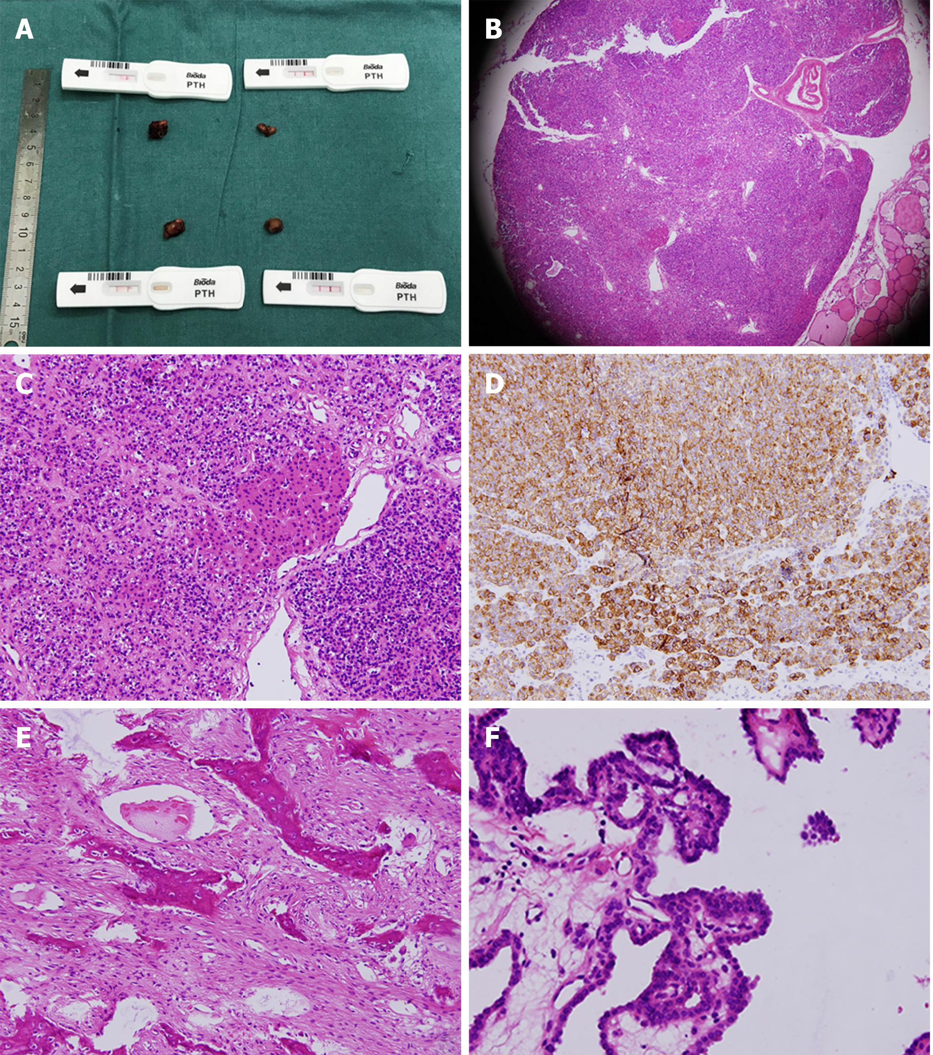
Figure 4 Histopathological examination of secondary hyperparathyroidism, mandibular biopsy, and papillary thyroid cancer.
A: Four parathyroid glands were confirmed by colloidal gold-based immunoassays during the operation; B and C: Histochemistry of the hyperplastic parathyroid gland by microscopy at 200× and 400×; D: Immunohistochemistry of the hyperplastic parathyroid gland by microscopy at 400×; E: Bone trabeculae could be seen in the fibroblast background with multinucleated megakaryocytes; F: Histochemistry of the papillary thyroid carcinoma by microscopy at 400×.
- Citation: Yu Y, Zhu CF, Fu X, Xu H. Sagliker syndrome: A case report of a rare manifestation of uncontrolled secondary hyperparathyroidism in chronic renal failure. World J Clin Cases 2019; 7(22): 3792-3799
- URL: https://www.wjgnet.com/2307-8960/full/v7/i22/3792.htm
- DOI: https://dx.doi.org/10.12998/wjcc.v7.i22.3792


