Copyright
©The Author(s) 2019.
World J Clin Cases. Nov 26, 2019; 7(22): 3778-3783
Published online Nov 26, 2019. doi: 10.12998/wjcc.v7.i22.3778
Published online Nov 26, 2019. doi: 10.12998/wjcc.v7.i22.3778
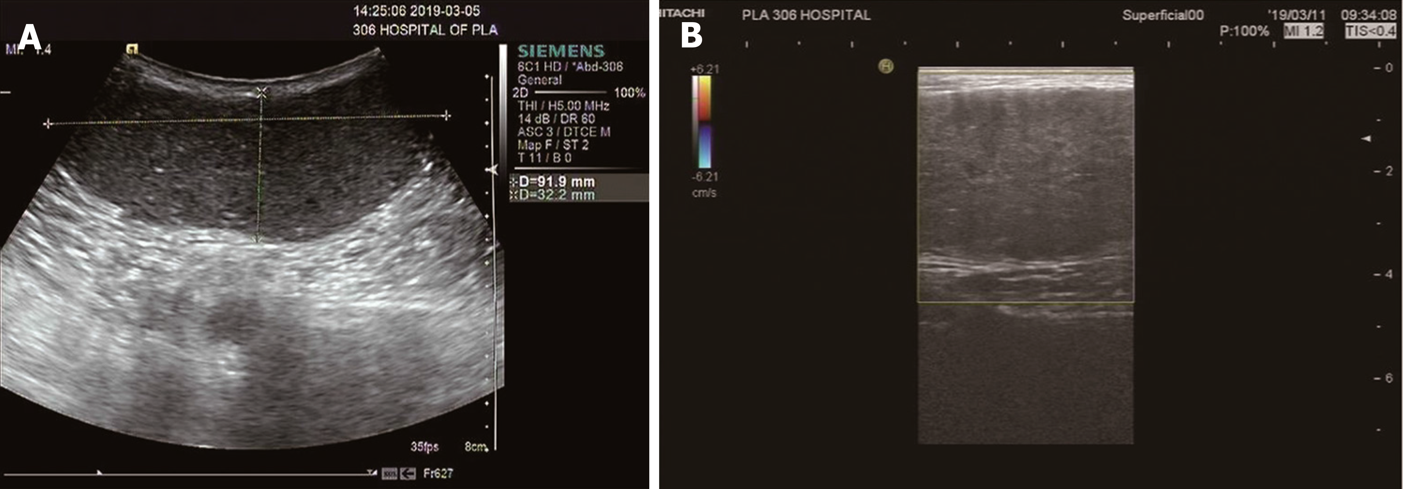
Figure 1 Ultrasound images.
A: Ultrasound image showing a hypoechoic mass, with the maximum range of 9.2 cm × 3.7 cm, clear margins, and regular morphology; B: Color Doppler flow image indicating that there was no obvious blood flow in the mass.
- Citation: Sun PM, Yang HM, Zhao Y, Yang JW, Yan HF, Liu JX, Sun HW, Cui Y. Contrast-enhanced computed tomography findings of a huge perianal epidermoid cyst: A case report. World J Clin Cases 2019; 7(22): 3778-3783
- URL: https://www.wjgnet.com/2307-8960/full/v7/i22/3778.htm
- DOI: https://dx.doi.org/10.12998/wjcc.v7.i22.3778


