Copyright
©The Author(s) 2019.
World J Clin Cases. Nov 26, 2019; 7(22): 3772-3777
Published online Nov 26, 2019. doi: 10.12998/wjcc.v7.i22.3772
Published online Nov 26, 2019. doi: 10.12998/wjcc.v7.i22.3772
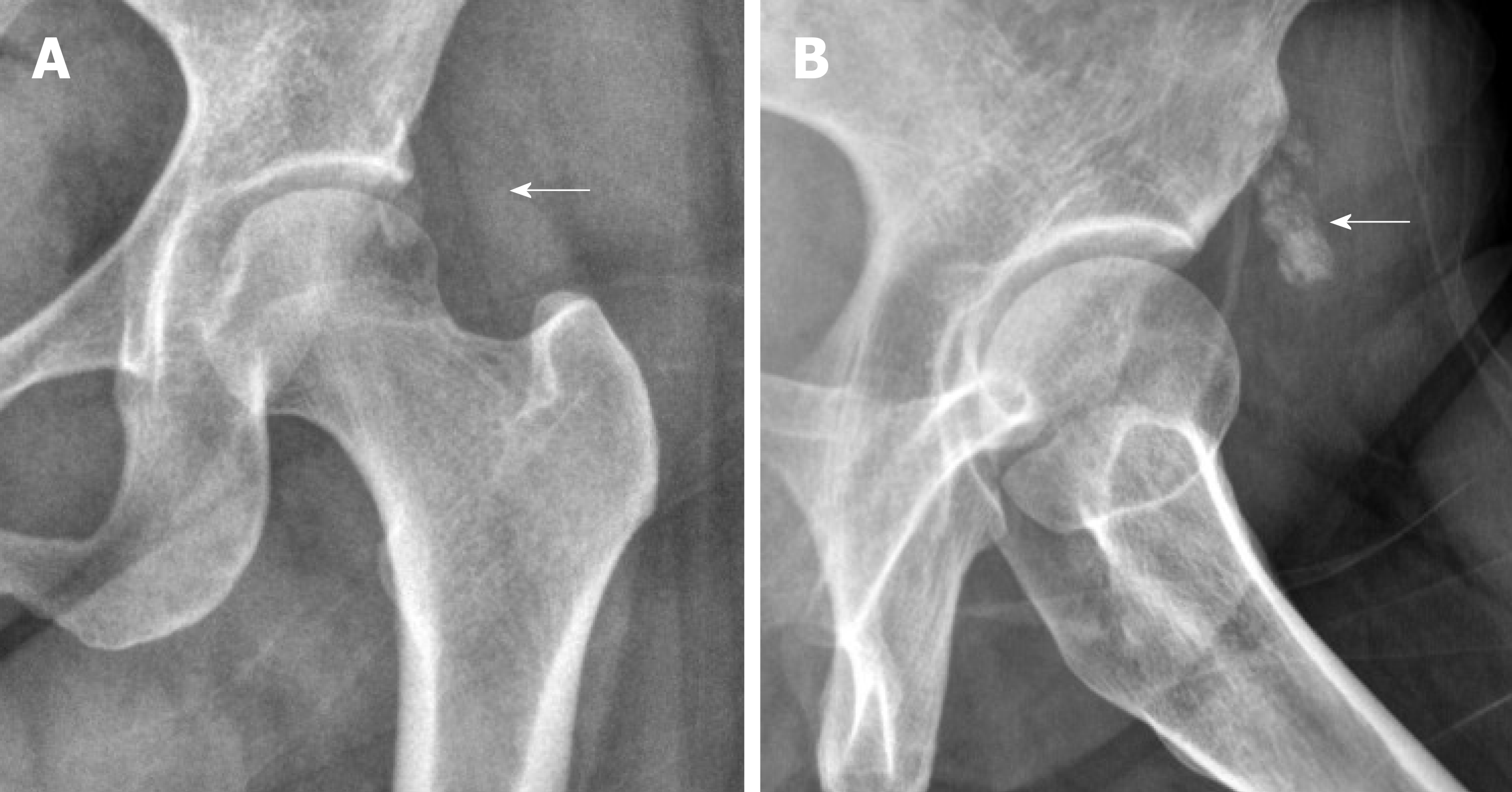
Figure 3 Simple radiography.
A: The size of calcification was almost not reduced in simple radiography at the final follow-up; B: The lesion (white arrow) was still clearly defined at lateral view.
- Citation: Lee CH, Oh MK, Yoo JI. Ultrasonographic evaluation of the effect of extracorporeal shock wave therapy on calcific tendinopathy of the rectus femoris tendon: A case report. World J Clin Cases 2019; 7(22): 3772-3777
- URL: https://www.wjgnet.com/2307-8960/full/v7/i22/3772.htm
- DOI: https://dx.doi.org/10.12998/wjcc.v7.i22.3772


