Copyright
©The Author(s) 2019.
World J Clin Cases. Nov 26, 2019; 7(22): 3698-3710
Published online Nov 26, 2019. doi: 10.12998/wjcc.v7.i22.3698
Published online Nov 26, 2019. doi: 10.12998/wjcc.v7.i22.3698
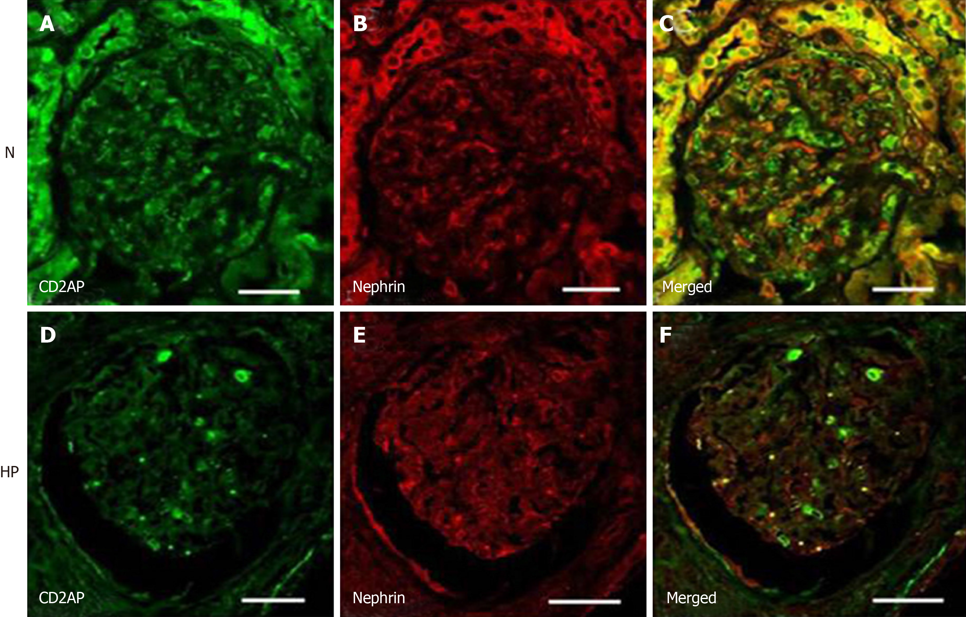
Figure 5 Confocal laser scanning micrographs show double immunofluorescence of nephrin–CD2-associated protein under normotensive and hypertensive conditions.
Under normotensive conditions, double immunofluorescence staining showed that CD2-associated protein (CD2AP) (A) and nephrin (B) partially colocalized along the basement membrane of glomeruli (C). Under hypertensive conditions, the immunoreactivity of CD2AP and nephrin decreased and stained intermittently (D-F). Scale bar = 20 μm. CD2AP: CD2-associated protein; N: Normotension; HP: Hypertension.
- Citation: Sun D, Wang JJ, Wang W, Wang J, Wang LN, Yao L, Sun YH, Li ZL. Human podocyte injury in the early course of hypertensive renal injury. World J Clin Cases 2019; 7(22): 3698-3710
- URL: https://www.wjgnet.com/2307-8960/full/v7/i22/3698.htm
- DOI: https://dx.doi.org/10.12998/wjcc.v7.i22.3698


