Copyright
©The Author(s) 2019.
World J Clin Cases. Nov 26, 2019; 7(22): 3698-3710
Published online Nov 26, 2019. doi: 10.12998/wjcc.v7.i22.3698
Published online Nov 26, 2019. doi: 10.12998/wjcc.v7.i22.3698
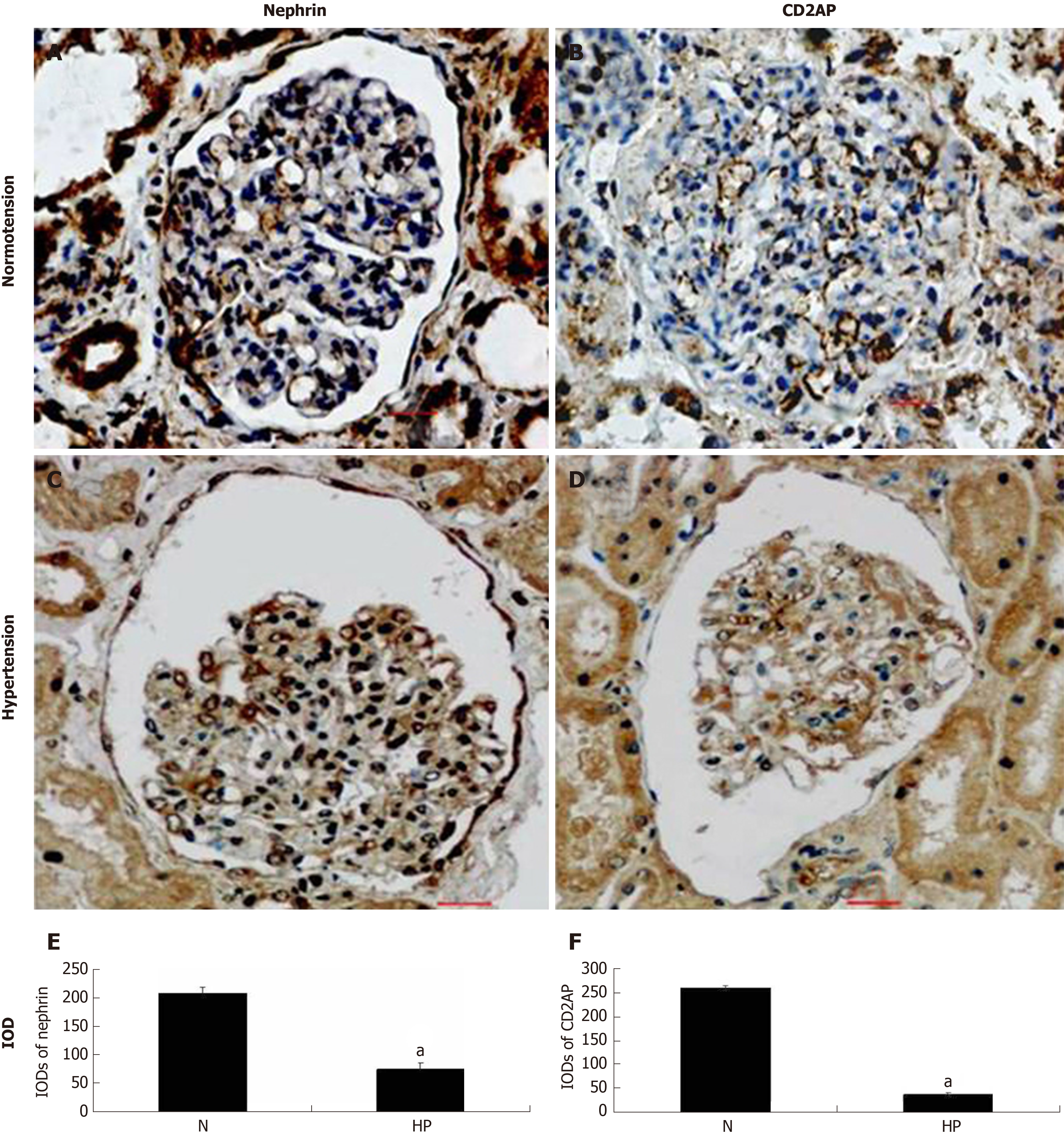
Figure 4 Expression of nephrin and CD2-associated protein in human renal tissue by immunohistochemical staining.
Tissues were treated with immersion-fixation methods under various hemodynamic conditions. A, C: The localization of nephrin; B, D: The localization of CD2-associated protein; E, F: The integral optical densities of each protein under various hemodynamic conditions. Immunohistochemical localization of both proteins in paraffin-embedded tissue is shown under normotensive (A, B) and hypertensive (C, D) conditions. Scale bar = 20 μm. Histogram shows integral optical density of each protein. aP < 0.05, Normotension vs Hypertension. N: Normotension; HP: Hypertension; IODs: Integral optical densities; CD2AP: CD2-associated protein.
- Citation: Sun D, Wang JJ, Wang W, Wang J, Wang LN, Yao L, Sun YH, Li ZL. Human podocyte injury in the early course of hypertensive renal injury. World J Clin Cases 2019; 7(22): 3698-3710
- URL: https://www.wjgnet.com/2307-8960/full/v7/i22/3698.htm
- DOI: https://dx.doi.org/10.12998/wjcc.v7.i22.3698


