Copyright
©The Author(s) 2019.
World J Clin Cases. Nov 6, 2019; 7(21): 3655-3661
Published online Nov 6, 2019. doi: 10.12998/wjcc.v7.i21.3655
Published online Nov 6, 2019. doi: 10.12998/wjcc.v7.i21.3655
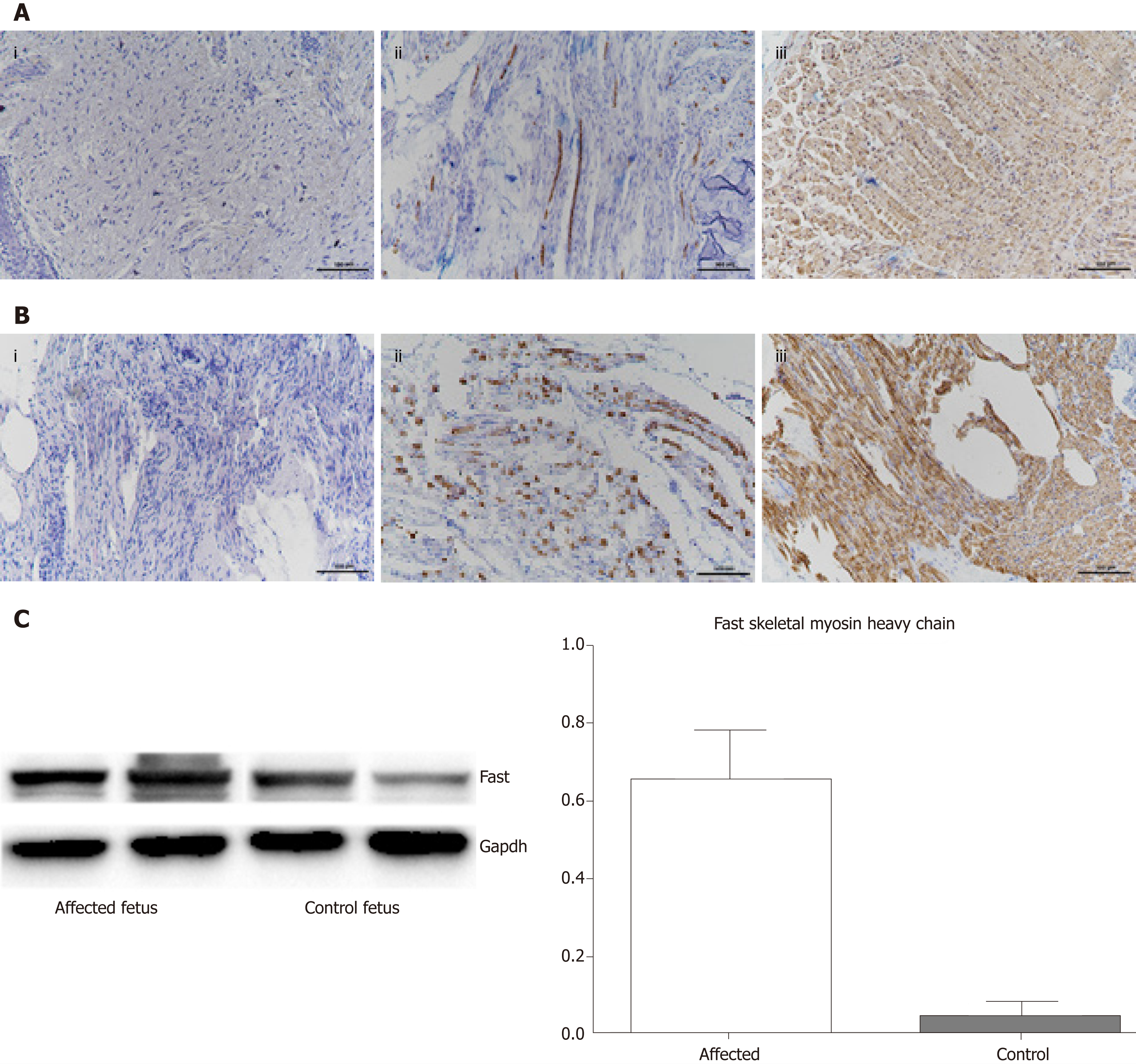
Figure 2 Histological, immunohistochemical, and Western blot findings.
A: Tissues from the affected fetus; A i: Hematoxylin and eosin staining showed a large variation in muscle fiber size, with many atrophic fibers and increased amounts of loose connective tissue; A ii: Immunohistochemistry with antibody against slow myosin demonstrated only a few scattered type 1 fibers; A iii: Immunohistochemistry with antibody against fast myosin demonstrated that the majority were muscle fibers; B: The results for a control fetus (dead fetus after a spontaneous abortion after the same number of gestational weeks); C: Western blot indicating that the amount of fast myosin heavy chain was significantly higher in the muscles of the affected fetus compared to the control fetus. The loading control is GAPDH. P < 0.05.
- Citation: Li N, Qiao C, Lv Y, Yang T, Liu H, Yu WQ, Liu CX. Compound heterozygous mutation of MUSK causing fetal akinesia deformation sequence syndrome: A case report. World J Clin Cases 2019; 7(21): 3655-3661
- URL: https://www.wjgnet.com/2307-8960/full/v7/i21/3655.htm
- DOI: https://dx.doi.org/10.12998/wjcc.v7.i21.3655


