Copyright
©The Author(s) 2019.
World J Clin Cases. Nov 6, 2019; 7(21): 3639-3648
Published online Nov 6, 2019. doi: 10.12998/wjcc.v7.i21.3639
Published online Nov 6, 2019. doi: 10.12998/wjcc.v7.i21.3639
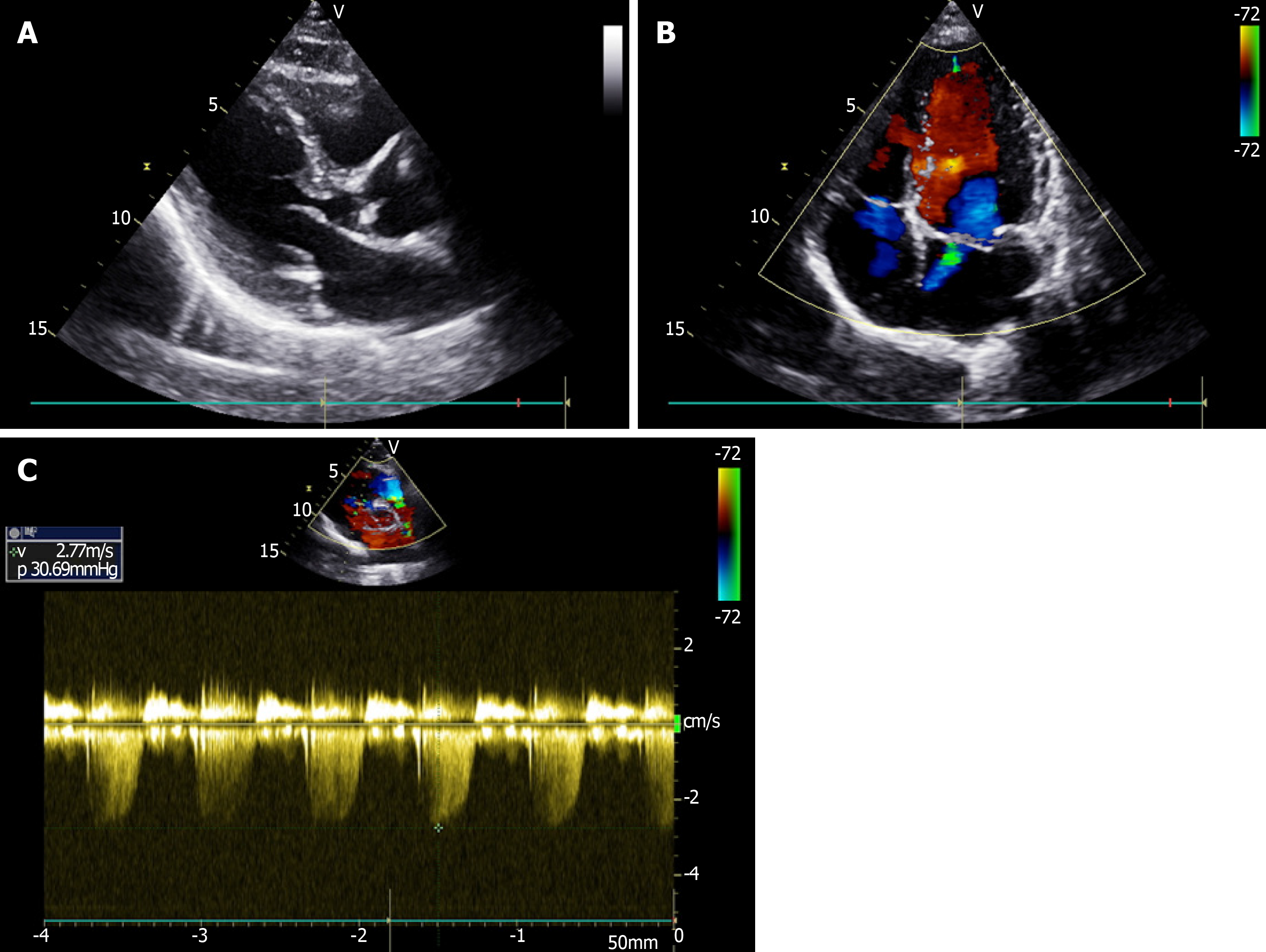
Figure 3 Echocardiography imaging examinations.
A: Showed calcification of the posterior mitral valve ring; B, C: Mild regurgitation of the mitral and tricuspid valves.
- Citation: Sun YY, Zhang GM, Zhang YB, Du X, Su ML. Bilateral common carotid artery common trunk with aberrant right subclavian artery combined with right subclavian steal syndrome: A case report. World J Clin Cases 2019; 7(21): 3639-3648
- URL: https://www.wjgnet.com/2307-8960/full/v7/i21/3639.htm
- DOI: https://dx.doi.org/10.12998/wjcc.v7.i21.3639


