Copyright
©The Author(s) 2019.
World J Clin Cases. Nov 6, 2019; 7(21): 3615-3621
Published online Nov 6, 2019. doi: 10.12998/wjcc.v7.i21.3615
Published online Nov 6, 2019. doi: 10.12998/wjcc.v7.i21.3615
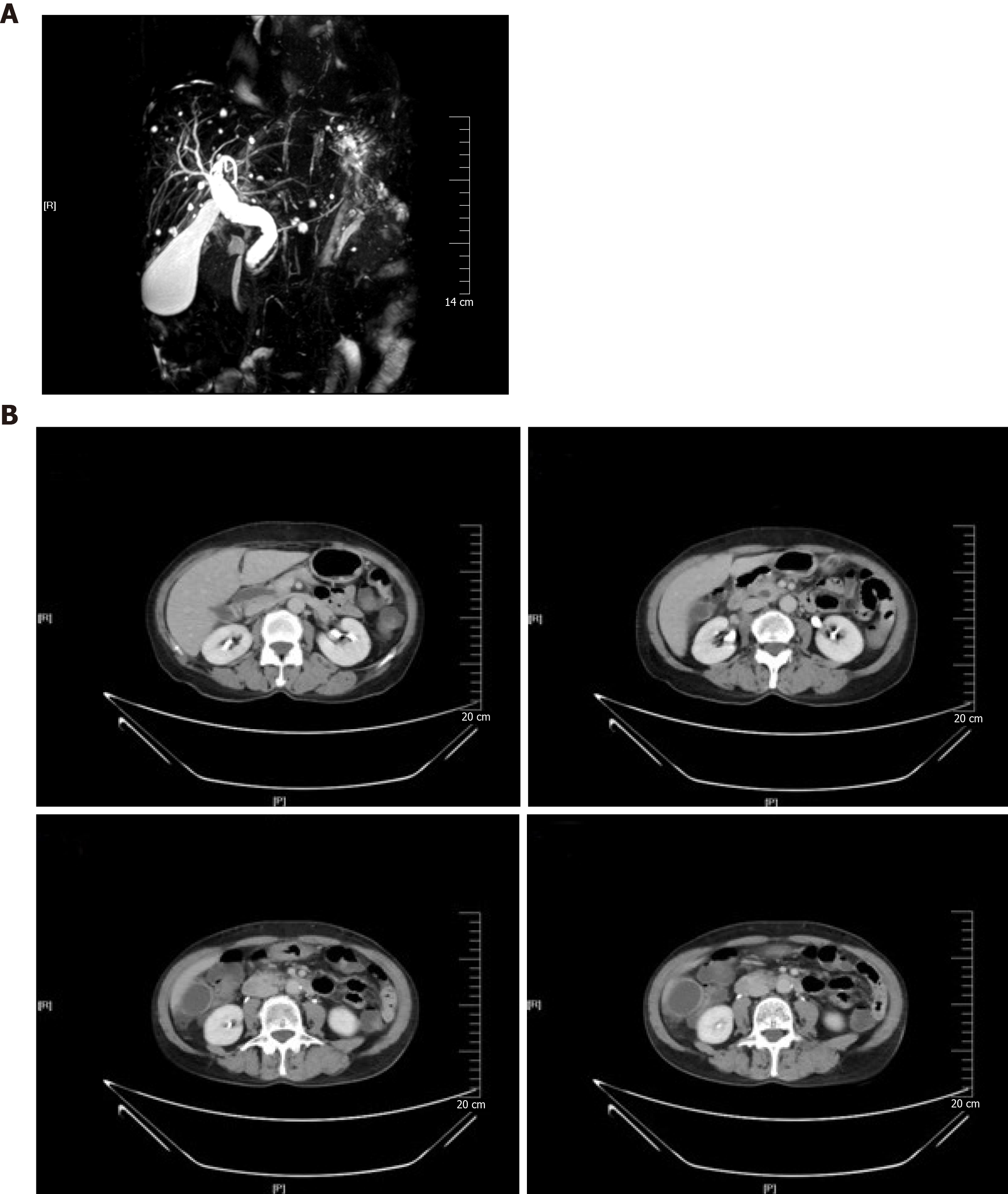
Figure 1 Magnetic resonance cholangiopancreatography and computed tomography images from our case.
A: Magnetic resonance cholangiopancreatography image showing proximal bile duct dilatation but ambiguous findings for evaluation of the distal common bile duct; B: Computed tomography images showing diffused dilatation of the extra-hepatic bile duct and significantly enhanced bile duct wall.
- Citation: Xu LM, Hu DM, Tang W, Wei SH, Chen W, Chen GQ. Adenomyoma of the distal common bile duct demonstrated by endoscopic ultrasound: A case report and review of the literature. World J Clin Cases 2019; 7(21): 3615-3621
- URL: https://www.wjgnet.com/2307-8960/full/v7/i21/3615.htm
- DOI: https://dx.doi.org/10.12998/wjcc.v7.i21.3615


