Copyright
©The Author(s) 2019.
World J Clin Cases. Nov 6, 2019; 7(21): 3595-3602
Published online Nov 6, 2019. doi: 10.12998/wjcc.v7.i21.3595
Published online Nov 6, 2019. doi: 10.12998/wjcc.v7.i21.3595
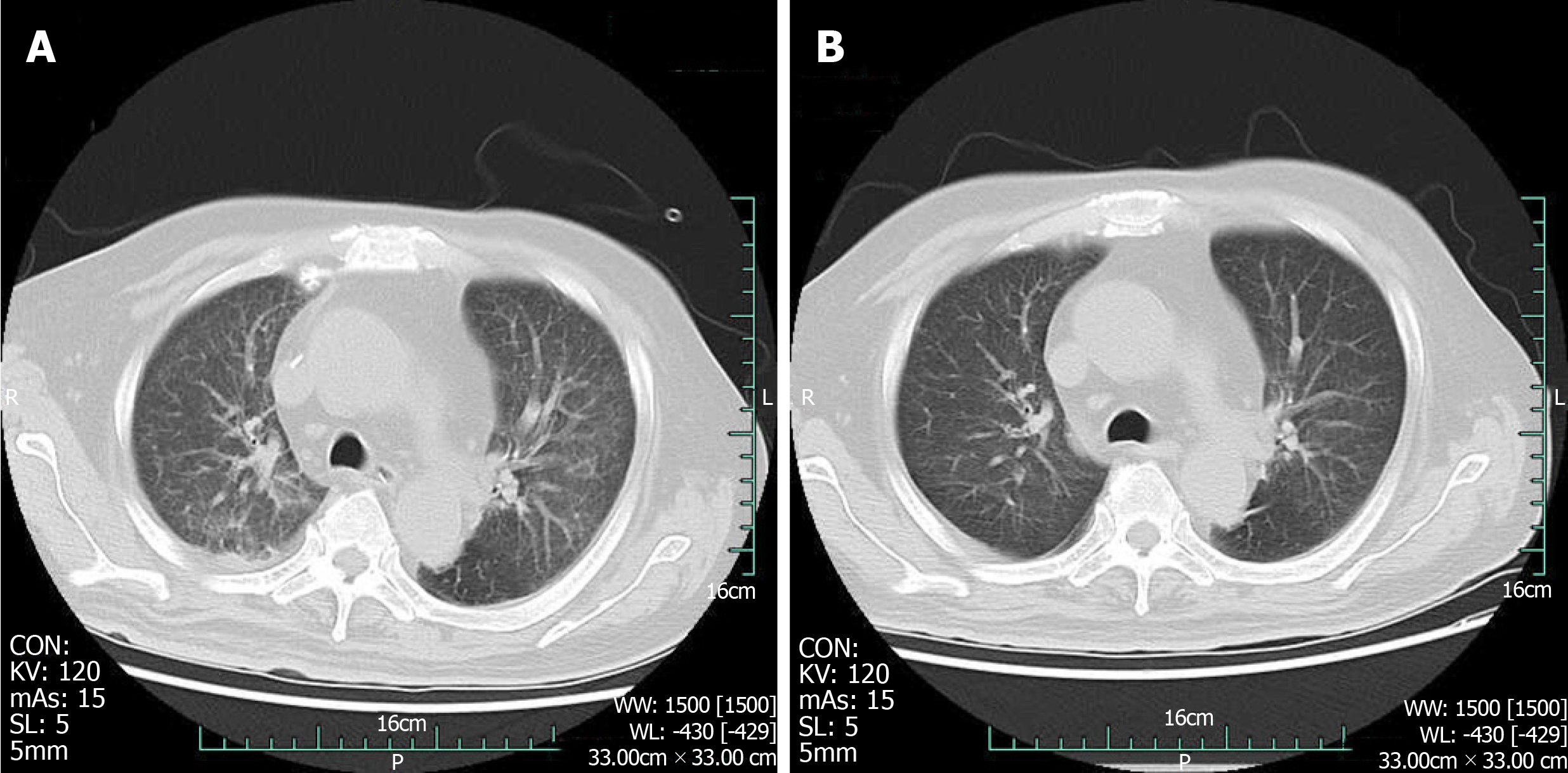
Figure 5 Computed tomography scan of the chest.
A: Acute bilateral pulmonary oedema and a small amount of pleural effusions at the base of both lungs; B: Bilateral pulmonary oedema and pleural fluid were absorbed.
- Citation: Xu LQ, Zhao XX, Wang PX, Yang J, Yang YM. Multidisciplinary treatment of a patient with necrotizing fasciitis caused by Staphylococcus aureus: A case report. World J Clin Cases 2019; 7(21): 3595-3602
- URL: https://www.wjgnet.com/2307-8960/full/v7/i21/3595.htm
- DOI: https://dx.doi.org/10.12998/wjcc.v7.i21.3595


