Copyright
©The Author(s) 2019.
World J Clin Cases. Nov 6, 2019; 7(21): 3562-3568
Published online Nov 6, 2019. doi: 10.12998/wjcc.v7.i21.3562
Published online Nov 6, 2019. doi: 10.12998/wjcc.v7.i21.3562
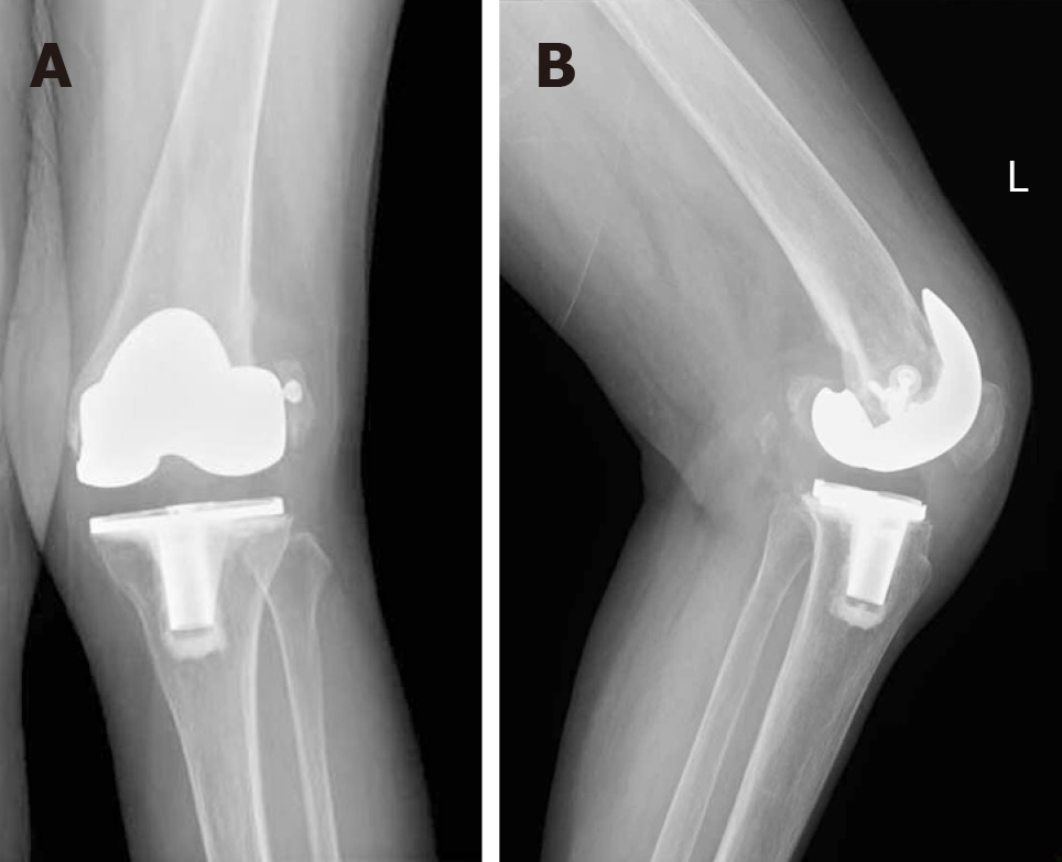
Figure 1 The radiographs of left knee at 6 months after primary total knee arthroplasty showed the signs of femoral component loosening and recurrent valgus deformity.
A: Anteroposterior radiograph; B: Lateral radiograph.
- Citation: Du YQ, Sun JY, Ni M, Zhou YG. Re-revision surgery for re-recurrent valgus deformity after revision total knee arthroplasty in a patient with a severe valgus deformity: A case report. World J Clin Cases 2019; 7(21): 3562-3568
- URL: https://www.wjgnet.com/2307-8960/full/v7/i21/3562.htm
- DOI: https://dx.doi.org/10.12998/wjcc.v7.i21.3562


