Copyright
©The Author(s) 2019.
World J Clin Cases. Nov 6, 2019; 7(21): 3553-3561
Published online Nov 6, 2019. doi: 10.12998/wjcc.v7.i21.3553
Published online Nov 6, 2019. doi: 10.12998/wjcc.v7.i21.3553
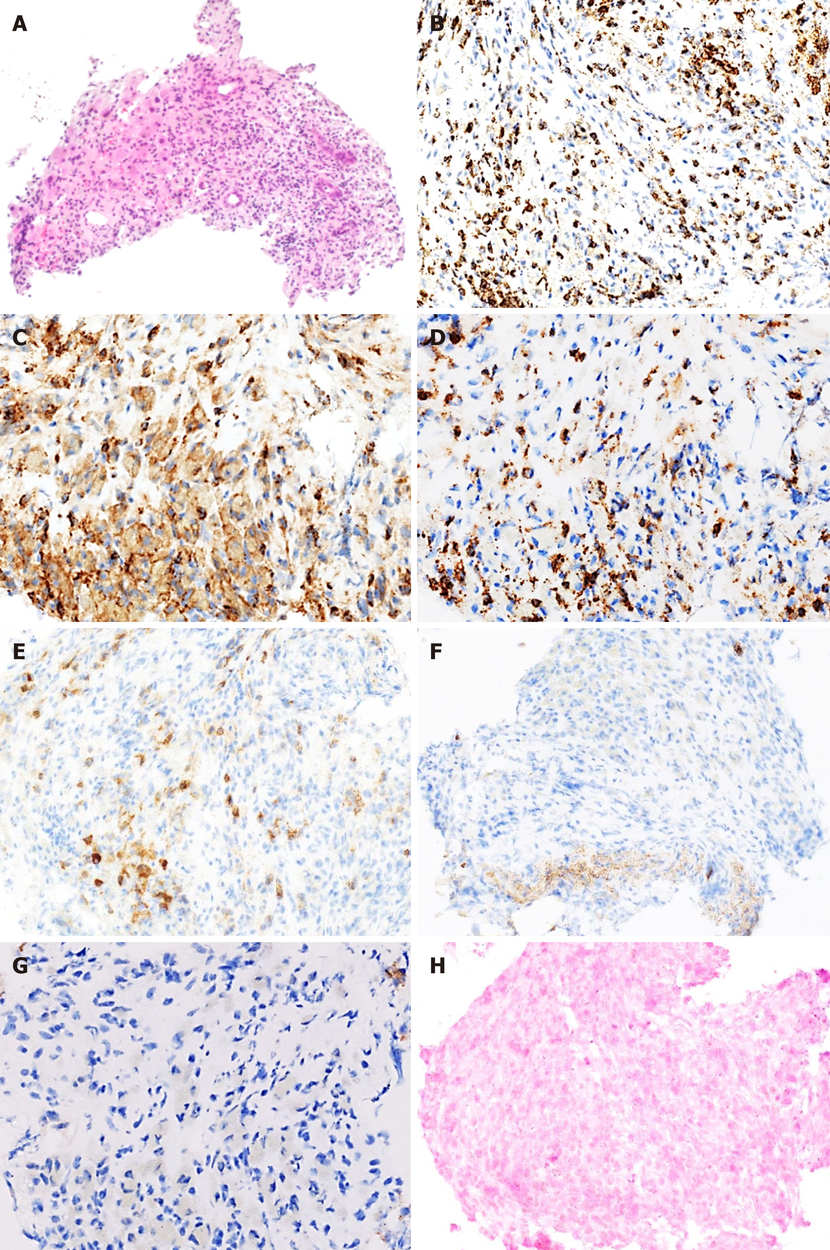
Figure 2 Histology of liver tumors in a patient with methotrexate-associated lymphoproliferative disorder.
HE staining (A, × 10) shows significant proliferation of lymphocytes in the liver, seen as masses that, on immunohistochemical staining; are positive for CD3 (B, × 20); CD4 (C, × 40); CD8 (D, × 40); and CD79a (E, × 20) but negative for CD20 (F, × 20) and CD56 (G, × 40). Staining for Epstein-Barr virus-encoded RNA (H, × 20) was negative.
- Citation: Mizusawa T, Kamimura K, Sato H, Suda T, Fukunari H, Hasegawa G, Shibata O, Morita S, Sakamaki A, Yokoyama J, Saito Y, Hori Y, Maruyama Y, Yoshimine F, Hoshi T, Morita S, Kanefuji T, Kobayashi M, Terai S. Methotrexate-related lymphoproliferative disorders in the liver: Case presentation and mini-review. World J Clin Cases 2019; 7(21): 3553-3561
- URL: https://www.wjgnet.com/2307-8960/full/v7/i21/3553.htm
- DOI: https://dx.doi.org/10.12998/wjcc.v7.i21.3553


