Copyright
©The Author(s) 2019.
World J Clin Cases. Nov 6, 2019; 7(21): 3524-3534
Published online Nov 6, 2019. doi: 10.12998/wjcc.v7.i21.3524
Published online Nov 6, 2019. doi: 10.12998/wjcc.v7.i21.3524
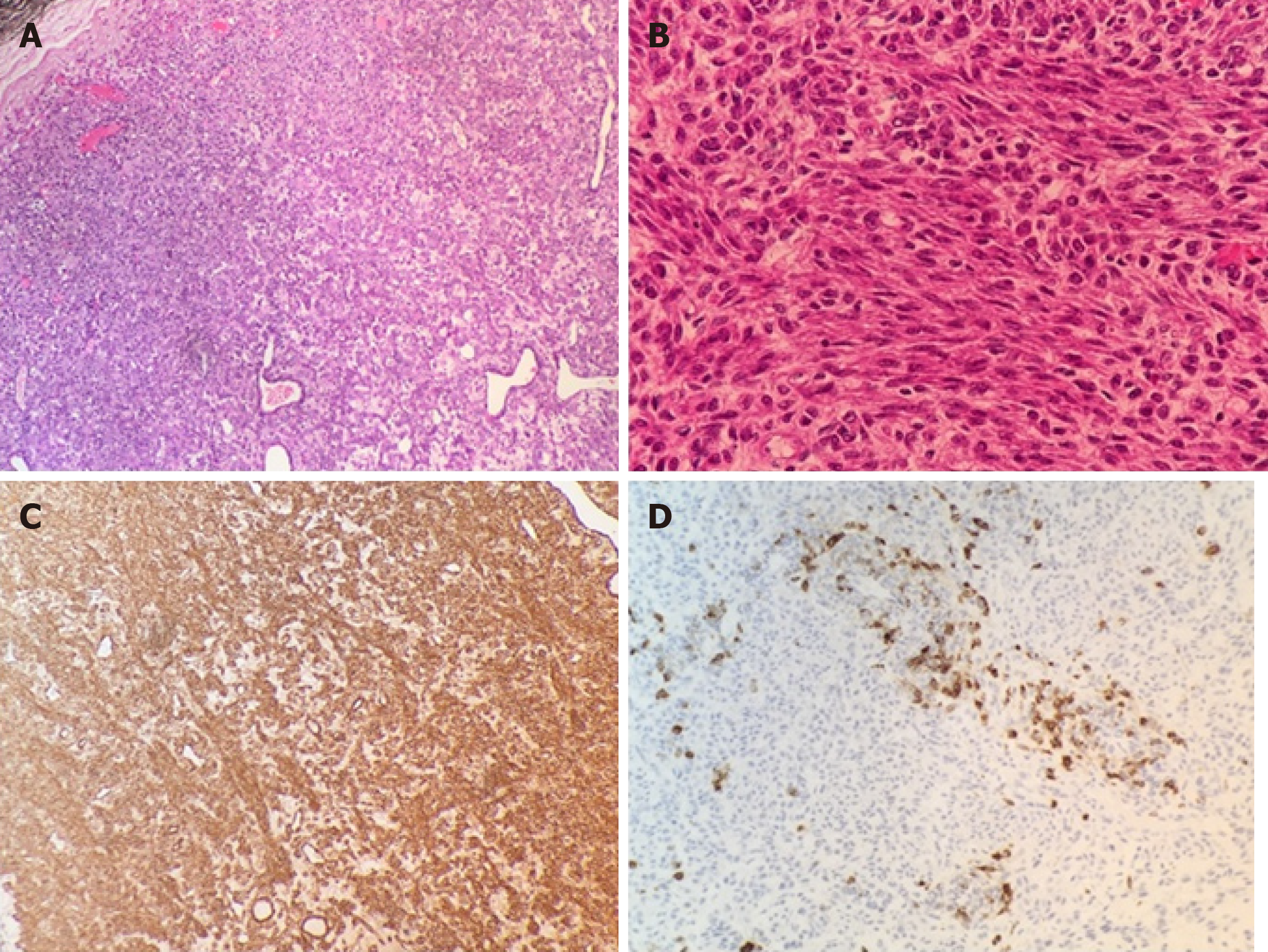
Figure 2 Histology and immunohistochemical analysis.
A: Encapsulated tumour composed of eosinophilic cells. Thin-walled and ectatic vessels are also apparent. Part of the tumour capsule is seen in the upper left of the picture (haematoxylin and eosin staining; magnification × 10). B: Polygonal or spindle cells, with eosinophilic cytoplasm and mildly atypical nuclei arranged in short bundles, around compressed, thin-walled vascular channels (haematoxylin and eosin staining; magnification × 40). C: Diffuse smooth muscle actin reactivity (magnification × 10). D: Focal human melanoma black-45 reactivity (magnification × 10).
- Citation: Touloumis Z, Giannakou N, Sioros C, Trigka A, Cheilakea M, Dimitriou N, Griniatsos J. Retroperitoneal perivascular epithelioid cell tumours: A case report and review of literature. World J Clin Cases 2019; 7(21): 3524-3534
- URL: https://www.wjgnet.com/2307-8960/full/v7/i21/3524.htm
- DOI: https://dx.doi.org/10.12998/wjcc.v7.i21.3524


