Copyright
©The Author(s) 2019.
World J Clin Cases. Oct 26, 2019; 7(20): 3377-3383
Published online Oct 26, 2019. doi: 10.12998/wjcc.v7.i20.3377
Published online Oct 26, 2019. doi: 10.12998/wjcc.v7.i20.3377
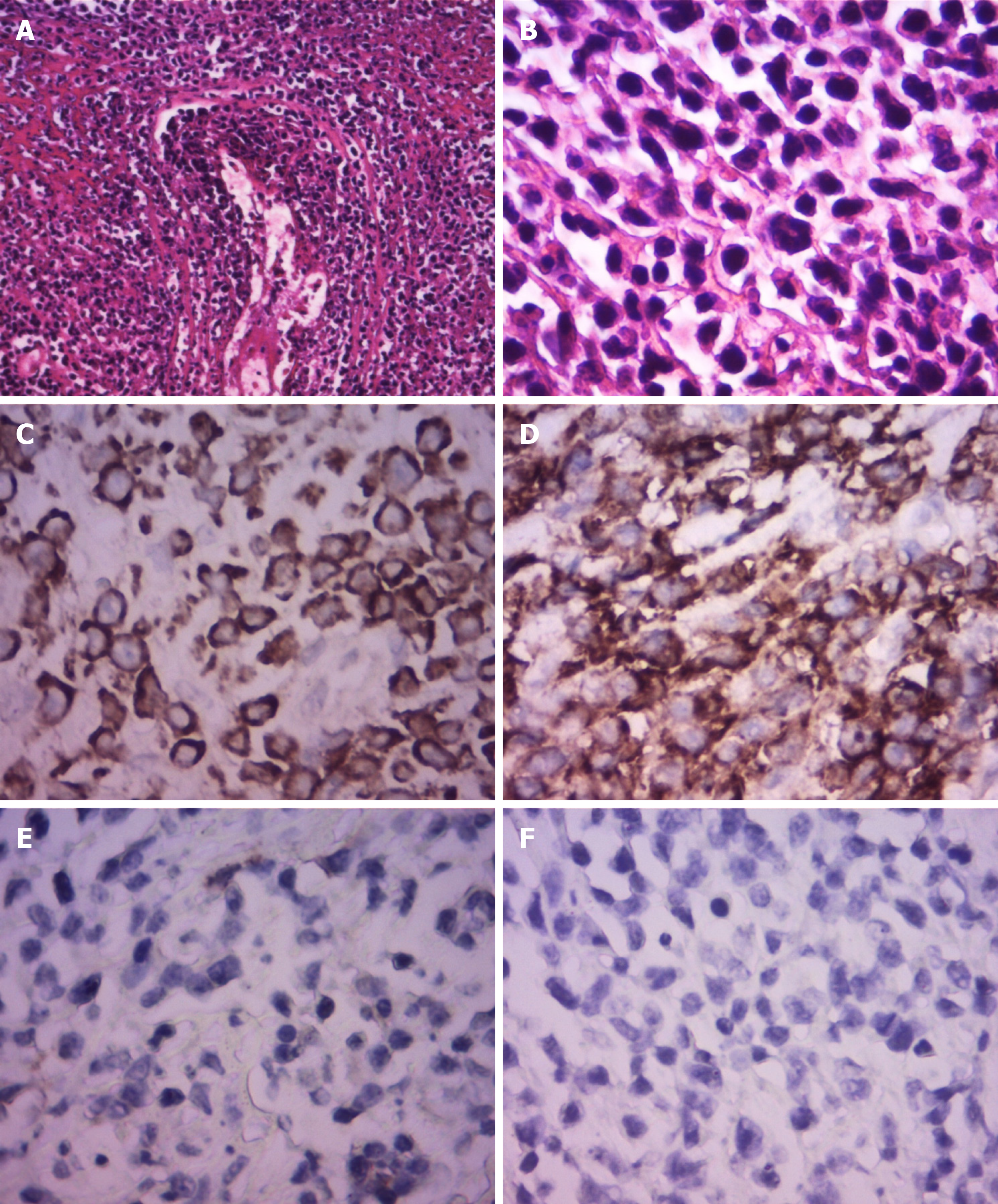
Figure 3 Pathological and immunohistological examination.
Immunohistochemistry showed the neoplastic cells were positive for CD2, CD3, CD10, CD30, Ki67, LCA, and Mum-1. The cells were negative for CD20, Bcl-6, Bcl-2, Pax-5, P63, PCK, CD56, P40, ALK-80, and EBER. Pathological examination: A: x 100; B: x 400; Immunohistological examination: C: CD3 (+), x 400; D: CD30 (+), x 400; E: CD56 (-), x 400; F: ALK-80 (-), x 400.
- Citation: Luo J, Jiang YH, Lei Z, Miao YL. Anaplastic lymphoma kinase-negative anaplastic large cell lymphoma masquerading as Behcet's disease: A case report and review of literature. World J Clin Cases 2019; 7(20): 3377-3383
- URL: https://www.wjgnet.com/2307-8960/full/v7/i20/3377.htm
- DOI: https://dx.doi.org/10.12998/wjcc.v7.i20.3377


