Copyright
©The Author(s) 2019.
World J Clin Cases. Oct 26, 2019; 7(20): 3377-3383
Published online Oct 26, 2019. doi: 10.12998/wjcc.v7.i20.3377
Published online Oct 26, 2019. doi: 10.12998/wjcc.v7.i20.3377
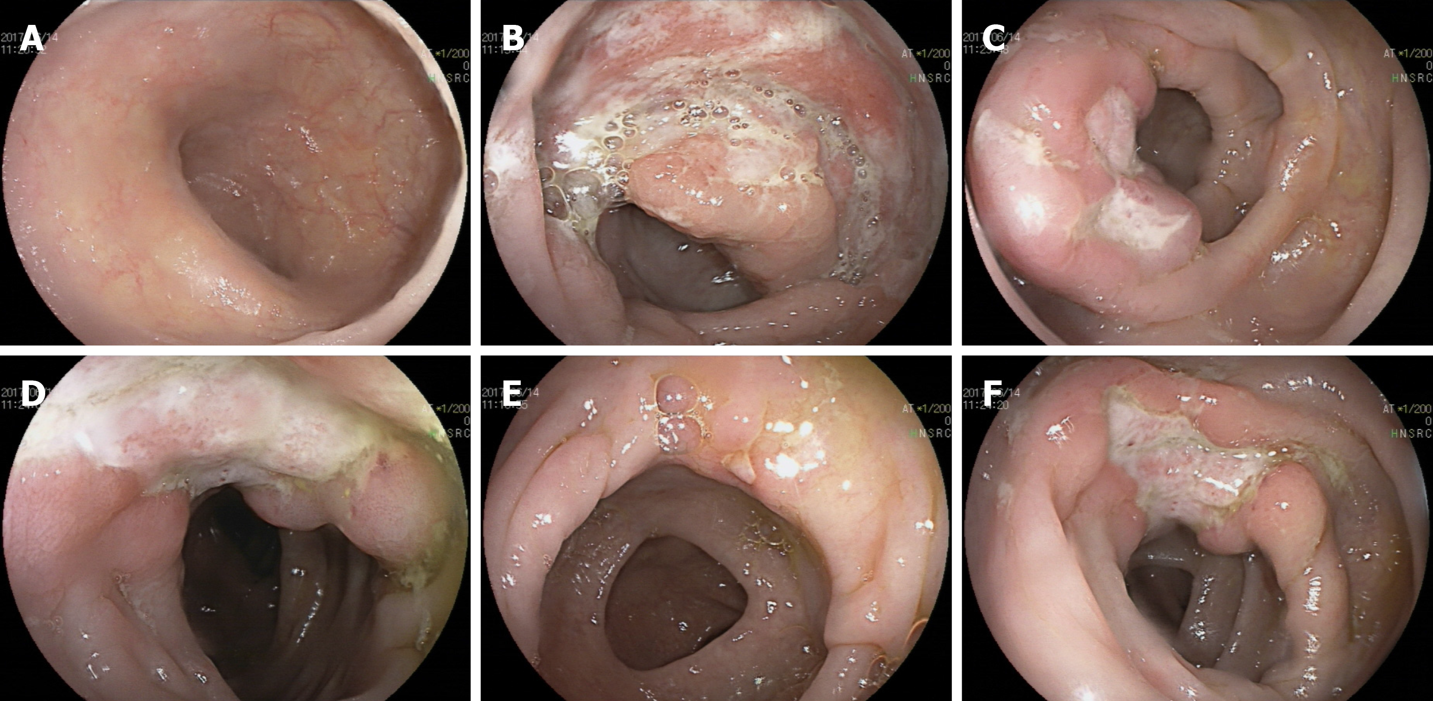
Figure 2 Colonoscopy.
The mucosa in the ileocecal region, ascending colon, transverse colon and descending colon were scattered with ulcers of varying sizes, all covered with white moss and surrounded by mucosal hyperemia and edema, especially in the ileocecal region:A: Distal ileum; B: Ileocecal region; C: Ascending colon; D: Transverse colon; E: Descending colon; F: Descending colon.
- Citation: Luo J, Jiang YH, Lei Z, Miao YL. Anaplastic lymphoma kinase-negative anaplastic large cell lymphoma masquerading as Behcet's disease: A case report and review of literature. World J Clin Cases 2019; 7(20): 3377-3383
- URL: https://www.wjgnet.com/2307-8960/full/v7/i20/3377.htm
- DOI: https://dx.doi.org/10.12998/wjcc.v7.i20.3377


