Copyright
©The Author(s) 2019.
World J Clin Cases. Oct 26, 2019; 7(20): 3377-3383
Published online Oct 26, 2019. doi: 10.12998/wjcc.v7.i20.3377
Published online Oct 26, 2019. doi: 10.12998/wjcc.v7.i20.3377
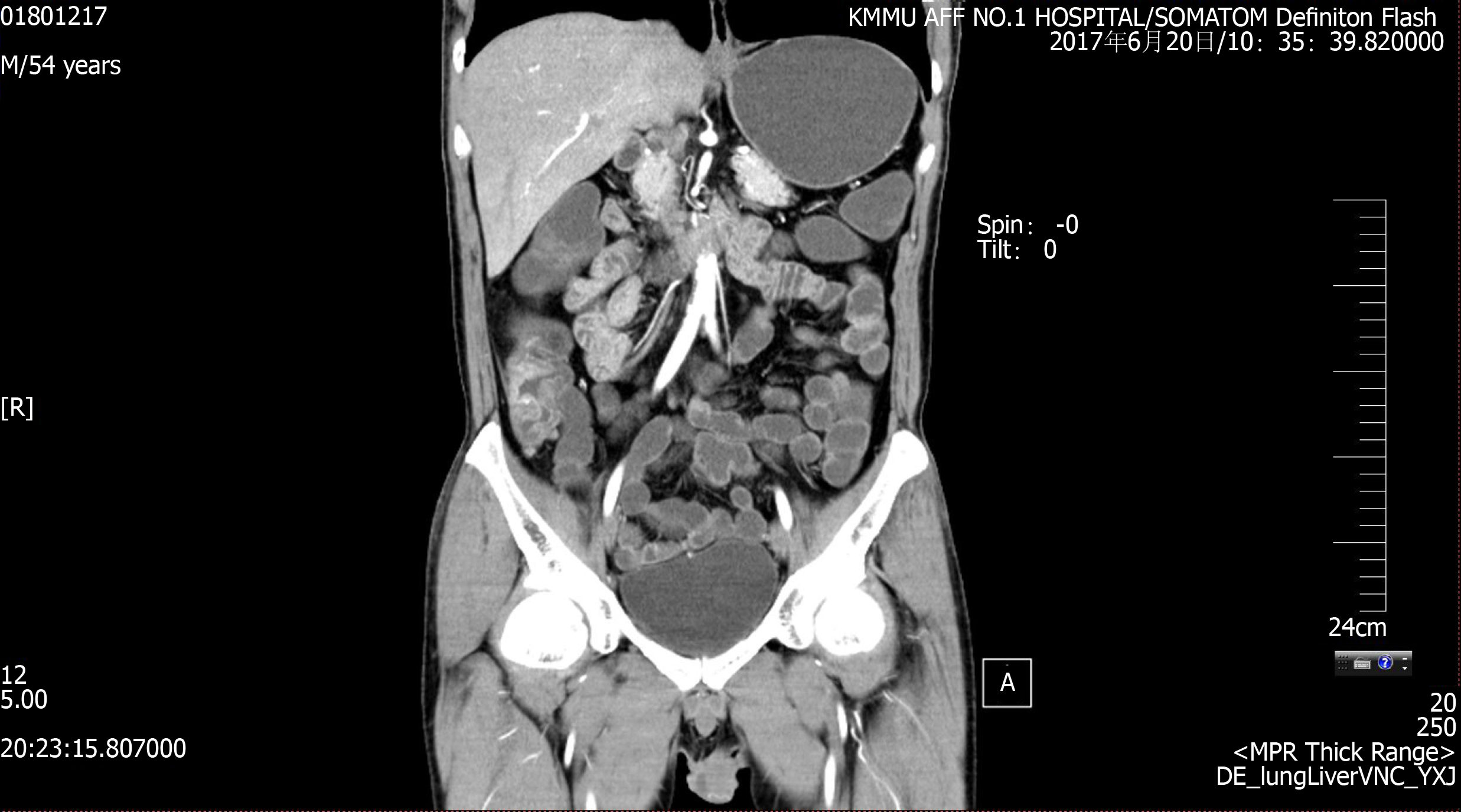
Figure 1 Computed tomography enterography.
Irregular thickening of the intestinal wall was observed in the cecum, ascending colon, transverse colon and descending colon, which also showed skip lesions. The thickest part of the intestinal wall was approximately 16 mm, and the intestinal wall in the arterial enhancement stage showed obvious enhancement (unclear hierarchical boundary); small vessels in the mesangial side were tortuous and dilated (locally formed as a "wooden comb"); and the surrounding fat was slightly blurred. No obvious thickening was observed in the small intestine wall.
- Citation: Luo J, Jiang YH, Lei Z, Miao YL. Anaplastic lymphoma kinase-negative anaplastic large cell lymphoma masquerading as Behcet's disease: A case report and review of literature. World J Clin Cases 2019; 7(20): 3377-3383
- URL: https://www.wjgnet.com/2307-8960/full/v7/i20/3377.htm
- DOI: https://dx.doi.org/10.12998/wjcc.v7.i20.3377


